Download
1 / 22
240 likes | 523 Views
Conception & Fetal Development Lecture 2. REVIEW Gametogenesis : production of gametes. Male gamete (sperm) produced in seminiferous tubules of testes during spermatogenesis. 200-600 mil.@ ejacula.

E N D
Conception & Fetal Development Lecture 2
REVIEW Gametogenesis: production of gametes. Male gamete (sperm) produced in seminiferous tubules of testes during spermatogenesis. 200-600 mil.@ ejacula. Female gamete (ovum) produced in graafian follicule of ovary during oogenesis. At birth, each ovary has 2 mil. immature oocytes, occurs 1st 5 mos. of development. Chromosomes divide (meiosis) from 46 → 23 before fertilization. 2 meiotic divisions in both sperm & ovum 1 spermatogonium >> 4 spermatids (approximately1000 sperm per second or ~ 30 billion/year) 1 oogonium >> 1 mature ovum & 3 polar bodies Ovum: 1st meiotic division completed before ovulation & 2nd meiotic division completed at fertilization.
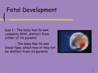
Conception: fertilization of sperm & ovum in ampulla [upper 1/3rd ] of fallopian tube. Now “zygote”. • ½ genetic material comes from each parent cell. Head of mature sperm contains chromosomes. • Zona pellucida (ovum) changes chemical composition so multiple sperm cannot enter. • Fertilized ovum begins mitotic cell division.
B. Cellular Multiplication • Zygote undergoes mitosis (cleavage) - rapid series of cell divisions. Forms morula; solid ball of cells. • Outer shell of cells with an attached inner group of cells forms, changing morula into “blastocyst”. • Blastocyst consists of inner cell mass and trophoblast. • Outer group of cells become membranes that nourish & protect inner group of cells (embryo). Blastocyst reaches uterus ~ 5th day. • Inside blastocyst, inner cell mass generates 3 major layers inside the sphere: ectoderm, mesoderm, endoderm. • Inner cell mass develops into fetus.
C. Implantation • Outer wall of blastocyst (trophoblast) attaches to endometrium (anterior or posterior fundal region) 7 - 9 days > fertilization. • Occurs 2-4 days > entering uterus. • Blastocyst receives nourishment via mother's bloodstream. • Embryo fully implanted by day 10. • During time between implantation & 8th week, cellular differentiation occurs.. (blood cells, kidney cells, nerve cells, etc.). • From 8th week until birth - “fetus”.
Yolk Sac • Attached to the embryo • Continuous with intestinal cavity of embryo • Supplied with blood vessels which transport nutritive yolk products to developing embryo • Degenerates by week 12 when placenta takes over
D. Placentation: ongoing process of fetal & maternal placental formation. • Begins immediately after implantation. • Endometrium now “decidua”. • 3 parts: basalis, capsularis, & vera. • Basalis unites with chorion to form maternal side of placenta. • Capsularis surrounds chorionic sac. • Decidua Vera is mucous membrane lining main cavity of pregnant uterus other than at site of implantation.
Pregnancy Hormones • Developing embryo begins to produce hCG (human chorionic gonadotropin) - enables corpus luteum to continue to secrete progesterone/estrogen. • In early pregnancy, steroid hormones are responsible for maintaining endometrium [uterus] rich with blood vessels so zygote can develop. • > 7th week shift to placental production of hormones begins. • By 12th week, hormone production entirely from placenta.
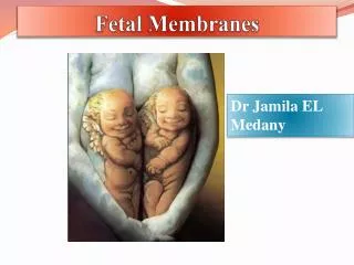
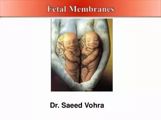
E. Fetal Membranes • Chorion: outer fetal membrane closest to uterine wall • Fingerlike projections “chorionic villi” form & invade endometruim; becomes fetal part of future placenta 2-3 weeks > fertilization. • Intervillous space: space between the chorionic villi where maternal blood circulates within placenta
Complex network of embryonic vessels allows diffusionof nutrients, oxygen, & wastes bet. mother & fetus. • Amnion: inner fetal membrane • Chorion & amnion fuse to become one membrane, amniotic sac. • Holds fetus & amniotic fluid.
F. Umbilical Cord: • 50-55 cm length; 2 cm diameter • 2 arteries & 1 vein – “AVA” • Made of Wharton’s jelly (gelatinous) • Blood flows thru cord @ rate of ~ 400 ml/min. • Vessels remain patent • Develops @ 5 wks.gestation; lifeline between placenta & fetus. • Carries waste & O2 poor blood away from fetus to placenta. • Carries O2 rich blood back to fetus.
G. Placenta Structure: 15-20 subdivisions “cotyledons”. • Fetal surface: smooth, shiny, covered by amnion. • Maternal surface: red, flesh-like, 15-20 subdivisions or cotyledons. • "Dirty Duncan" and "Shiny Schultz" Physiology: • Function: transport mechanism between mom & fetus. • Lifespan depends on O2 consumption. • Function depends on maternal circulation. • Circulation best when mother in lateral position.
Functions of Placenta • Receives maternal 02 via diffusion. • Produces all hormones to sustain endometrium thus the pregnancy (HPL, estrogen, progesterone, relaxin) • Supplies fetus with CHO, water, fats, proteins, minerals & inorganic salts. • Carries end products of fetal metabolism into maternal circulation for excretion. • Transfers passive immunity via maternal antibodies.
H. Amniotic Fluid • 800-1200ml. Clear, yellow fluid • Contains albumin, lanugo & urea. • Replaced every 3 hours; swallowed by fetus. Functions • Prevents heat loss; preserves constant fetal body temperatures. • Cushions fetus. • Acts as excretion – collection system. • Facilitates fetal growth & development
First Trimester - fetus most susceptible to damage from external sources including: • teratogens (causing birth defects … ie. alcohol, some Rx & recreational drugs) • infections (ie. rubella or cytomegalovirus) • radiation (x-rays, radiation therapy, or accidental exposure to radiation) • nutritional deficiencies
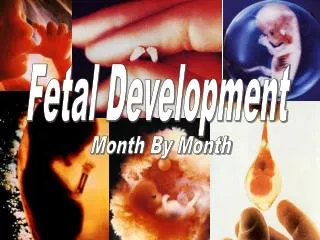
Summary of Fetal Development • 4th wk: Fetal heart begins to beat. (smaller than grain of rice). • 8 wks: All body organs formed. Weighs less than grape. 1/3 baby is head. Arms & legs are tiny buds. L: 30mm ( 1in.) Wt. = 2 g. • 12 wks: Fetal heart tones heard by Doppler. Baby can kick. Sex organs formed. Fingers & toes developed. Eyelids fused. L: 4 in or 11cm. W: 45 g.
Fetal Development • 16 wks: Sex can be seen. Thin; looks like baby. Uterus size of grapefruit. Fetus as large as orange. Starting to suck/swallow. Nails on fingers/toes. L: 5 in or 15 cm. W: 200 g. Actively swallows amniotic fluid. Lanugo forms. • 20 wks: Heartbeat heard with fetoscope. Develops regular schedule: sleeping, sucks thumb, kicking. Hands grasp.Vernix caseosa begins to form. Assumes favorite position in utero. Lanugo keeps oil on skin. L: 25 cm. W: 400 g. + fetal movement (quickening)
Fetal Development • 24 wks: L: 30 cm. W: 750 g. Weighs about 1.3 lb. Increased activity. Respiratory movement begins. Skin is thin. No fat. Regular sleep time. • 28 wks: Eyes open & close. Baby makes breathing motions. Surfactant begins forming. Testes descend. More fat forms. Can hiccup, cry, hear your voice. 14” long; 2.2 lbs. Baby 2/3rd final size. L: 35 cm. W: 1200g.
Fetal Development • 32 weeks: More subcutaneous fat laid down. Appears less red & wrinkled; 14” long; 4 lbs. • brain growing. Lungs immature. Gains 1/2 lb/wk. L:35-38 cm. W: 2000 g. Skin pink; covered with vernix caseosa; lanugo begins to disappear. Braxton Hicks are felt. • 38-40 wks: Full term @ beg. of 37th wk. Fills uterus. Gets IgA ab from mother. Gains 2 ½ lbs; mostly fat. L: 48-52 cm. or 20” long. W: 3000-4000g, or ~ 8 lbs.
Fetal Circulation • Placenta [O2 rich blood >> maternal circulation] • Enters Umbilical vein (O2 rich blood) • ductus venous (2/3rd bypasses liver) • hepatic vein (small amt. blood flow) • Enters inferior vena cava [IVC] • 30-35% enters R. atrium >> passes through foramen ovale (shunt in fetal heart) >> L. Atrium >> L.Ventricle >> Ascending aorta to Head & upper extremities (to oxygenate where needed most)
65-70%of blood in Right atrium mixes with O2 poor blood returning from SVC >> Right Ventricle >> enters ductus arteriosis to be shunted away from lungs >> enters descending aorta >> lower extremities/trunk • Only ~ 8 % enters pulmonary arterial bed through right & left pulmonary arteries and returns from lungs to left atrium via 4 pulmonary veins. • Eventually all O2 poor blood leaves thru aorta >> 2 umbilical arteries >> placenta to re-oxygenate.
Fetal circulation: Preferential shunting of blood with highest O2 saturation to L side of heart. Assures adequate oxygenated blood flow to coronary & cerebral circulations—tissues with greatest need. • Circulation > birth: With first breaths, larger amount of blood sent to lungs to pick up O2. Ductus arteriosus no longer needed; begins to wither & close off in 1-2 days. PDA may be heard 1st 24-48 hours of life. • Circulation in lungs ^ & more blood flows into L atrium. This ^ pressure causes foramen ovale to close & blood circulates normally. • Umbilical arteries/vein degenerate. Shunts & vessels > ligaments & supporting structures. http://www.indiana.edu/~anat550/cvanim/fetcirc/fetcirc.html