Download
1 / 1
10 likes | 138 Views
Changes in Heart Rate Volatility In A Murine Model Of Sepsis Goel N, Skaf J, Guglielmi M, Foley B, Zanotti S, Parrillo JE, Hollenberg SM Cardiology and Critical Care, Cooper University Hospital, Camden, NJ. Background

E N D
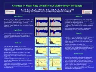
Changes in Heart Rate Volatility In A Murine Model Of Sepsis Goel N, Skaf J, Guglielmi M, Foley B,Zanotti S, Parrillo JE, Hollenberg SM Cardiology and Critical Care, Cooper University Hospital, Camden, NJ Background • Nonlinear analysis of hemodynamic parameters such as Heart Rate Variability (HRV) may provide insights not available from standard linear measures • Power spectral analysis of HRV is commonly used. However, challenges arise from artifacts and dense data capture. Hypothesis • Sepsis will be associated with perturbations in Heart Rate Volatility (standard deviation variability), a means of assessing HRV that minimizes artifact-induced error. Methods • C57/Bl6 mice (8-12 weeks, 20 g., n=24) • Radiotelemeters for hemodynamic measurements in awake animals were implanted in the ascending aorta via the carotid artery. • Animals were allowed to recover for 5 to 7 days. • Baseline data was obtained for 24 hours. • Sepsis was induced by cecal ligation and puncture (CLP, n=20). • Controls received sham-operation (SO, n = 4). • Animals were resuscitated with fluids and antibiotics every 6 hours. • Heart Rate (HR) was calculated from blood pressure waveforms obtained from radiotelemeters. • HR standard deviations (SD) were calculated on each 5 minute interval. Methods • For each animal, SD histograms were constructed and the cutoff that represented the lowest 5% was calculated for the baseline period. • The percentage of low SD’s (representing low HRV) in the entire experimental period was defined by this cutoff. • A time course was generated by calculating the percentage of low HRV over 4 hour intervals. Results • Animals in the control group had low HRV detected in 1.5% of all intervals (p =NS versus baseline) • Animals in the septic group had low HRV in 38.72% of intervals post-CLP (p<0.01 versus baseline and versus controls) • Mortality in the septic group was 60%. • Survivors and nonsurvivors had a similar decrease in HR volatility early, with partial recovery, but then HRV responses diverged, with normalization in survivors, and further perturbation in non-survivors. Conclusions • Analysis of Heart Rate Volatility is less demanding, more intuitive and less susceptible to artifact as a means of measuring HRV than spectral analysis. • We have shown dramatic differences between septic and control animals in a clinically relevant murine model of sepsis using these techniques. • Extrapolation of this methodology to critically ill patients has the potential to provide novel markers of hemodynamic decompensation.