Download
1 / 26
260 likes | 428 Views
3-D + Medical Imaging. Neb Duric 3-D ESO workshop June 12, 2008. Medical Imaging Cross-sectional views in the fundamental planes. Coronal. Sagittal. Transverse. Visible Human Project. Common devices Magnetic Resonance Imaging X-ray Computed Tomography

E N D
3-D+ Medical Imaging Neb Duric 3-D ESO workshop June 12, 2008
Medical Imaging Cross-sectional views in the fundamental planes. Coronal Sagittal Transverse Visible Human Project
Common devices Magnetic Resonance Imaging X-ray Computed Tomography Positron Emission Tomography Ultrasound Tomography Common applications Screening Diagnosis Therapy guidance Treatment monitoring Tomographic Imaging
Image Cubes CT Scans of Chest
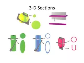
Projections of Image Cube coronal sagittal transverse
Volume Imaging Kai Uwe Barthel (k.barthel at fhtw-berlin.de)Internationale MedieninformatikBerlin, Germany
3-D + CharacterizationFusion Imaging Pretreatment 6 h post 18 d post 24 h post MRI Functional Imaging
Visualizing Risk PET/CT Functional Imaging
Confidential Acoustic Tomography
The Team Neb Duric1,2, Ph.D., Peter Littrup1,2, M.D., Cuiping Li1,2, Ph. D., Carri Glide1,2, Ph.D., Lisa Bey-Knight1,2, R.N., Yang Xu1,2,Ph.D., Steve Schmidt1,2, Olsi Rama1,2, Jason Shen1,2, PhD., Anthony Shields, M.D., Ph.D., Ding Wang, M.D., Ph.D., Lou Poulo3, M.Sc., Terry Kling4, Alex Babkin5, Ph.D.,Roman Pevzner5, Ph.D.,Lianjie Huang6, Ph.D., Francesco Simonetti7, Ph.D.,Ivana Jovanovic8 , Ph.D., Nicole Ruiter9, Ph.D., Frank Natterer10, Ph.D. 1Barbara Ann Karmanos Cancer Institute, Detroit, MI 2Wayne State University, Detroit, MI 3Analogic Corp, Boston, MA 4Sound Technology, State College, PA 5Groupvelocity LLC, Albuquerque, NM 6Los Alamos National Laboratory, Los Alamos, NM 7Imperial College, London, England 8Ecole Polytechnique Federale de Lausanne, Switzerland 9Forschungszentrum Karlsruhe,Karlsruhe, Germany 10University of Muenster, Muenster, Germany
Prototype ScannerImaging Tank and Ring Array Duric et al, Medical Physics Vol 34 (2), pp. 773-785, 2007 • In plane resolution: 0.5 – 2 mm • Out of plane resolution: 4 mm • 2 MHz operating frequency
Scan Method Reflection Sound Speed Attenuation
Thresholding and Segmentation Reflection Sound Speed Attenuation + + = = = Reflection Sound Speed Fusion Attenuation
Y X Z Volumetric 3-D / 4-D+ Imaging Operator independent imaging of entire breast volume. No compression or ionization. Exam time < 1 minute. Time Time
ResultsChemotherapy patients Reflection image Strong sound speed enhancement Strong attenuation Attenuation (blue) and sound speed (red) added Sound speed added (red)
ResultsQuantifying Breast Cancer Risk Santen, R J, Mansel, R. Benign breast disorders. N Engl J Med, 2005. 353(3): p. 275-85. • Known correlation between breast density and cancer risk • Currently measured by mammography
Our approachThresholding and clustering Ultrasound Percent Density (USPD) Sound Speed Tomogram Glide, C., Duric, N., Littrup, P. A Novel Approach to Evaluating Breast Density Utilizing Ultrasound Tomography. Medical Physics. Vol. 34 (2), pp. 744-753. Feb. 2007. Segmented Mammogram Segmented • Our study has established equivalence of sound speed and mammographic density potential for ultrasonic measure of breast density Potential Application: Chemoprevention and risk screening
SummarySimilarities and Differences Medical Astronomical Near Field Far Field Interactive Passive Super-resolution* Diffraction limited Structure noise Thermal noise Screening Surveys Diagnostic Targeted Imaging Characterization Spectral analysis Functional Kinematical ROC analysis ????? *Simonetti, Huang, Duric, Rama. Imaging beyond the Born approximation: An experimental investigation with an ultrasonic ring array. Physical Review E 76, 036601 (2007)
NGC 5055 Medical style Astronomical style Walter and Leroy
Acknowledgments • We acknowledge support from following grants: • Sponsor: NCI - Avon, • Improvements in Ultrasound Tomography for Characterization of Breast Masses: Duration: 12/06 – 11//07 • Sponsor: MEDC • Clinical Validation of CURE: A Novel Technology for Improved Breast Cancer Diagnosis: Duration: 02/07 – 05/09 • Komen Foundation grant. • Sound Speed as a Possible Predictor of Breast Cancer Risk.: Duration: 09/07-08/09. • Sponsor: Susan G. Komen foundation • Further reading: Duric, N., Littrup, P., Poulo, L., Babkin, A., Pevzner, R., Holsapple, E., Rama, O., Glide, C. Detection of Breast Cancer With Ultrasound Tomography: First Results with the Computerized Ultrasound Risk Evaluation (C.U.R.E) Prototype.Medical Physics Vol 34 (2), pp. 773-785. Feb 2007.
Structure noise and filtering Reflection Image Glandular Tissue defined largely by non-specular reflections Fibrous Stroma defined by specular reflections Measures Fibrous Stroma And Glandular tissue