Download
1 / 23
240 likes | 470 Views
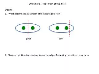
C. Cytokinesis. Usually occurs in the middle of the cell Exception: Sporulation and in Caulobacter Requires invagination of cell membrane, cell wall and outer membrane in Gm neg Form septum (membrane and wall) between cells. C. Cytokinesis. Fig 9.6 Microbe.

E N D
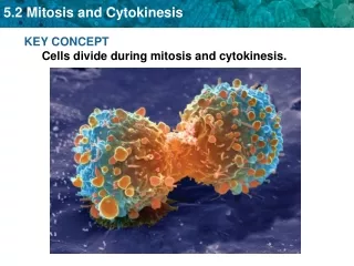
C. Cytokinesis • Usually occurs in the middle of the cell • Exception: Sporulation and in Caulobacter • Requires invagination of cell membrane, cell wall and outer membrane in Gm neg • Form septum (membrane and wall) between cells
C. Cytokinesis Fig 9.6 Microbe A. E. coli - Gram negative – • Constriction occurs in middle with invagination of inner membrane, cell wall and outer membrane, Septation(wall forming between) and separation occur together Fig 2.6 White Physiology and Biochem. of Prokaryotes
C. Cytokinesis • Bacillus - Gram positive – • Little constriction until end • septum(membrane) forms before separation Fig 9.7 Microbe Fig 2.6 White Physiology and Biochem. of Prokaryotes
C. Cytokinesis • Mechanisms not totally understood • Have identified factors involved • Many identified by temperature sensitive mutations that prevent cell division at non-permissive temperature • Cell division is an essential process • Isolate mutants that only stop dividing at a high temperature • Fts for filamentous temperature sensitive. Fig 9.10 Microbe
C. Cytokinesis Important factors • FtsZ – • Homolog of tubulin (protein that forms microtubules in eukaryotes) • Form filaments on inside of cell membrane • A bundle of filaments form a ring at cleavage site • ring gets smaller- causes contriction at cleavage site
C. Cytokinesis • Note how FtsZ localization changes during cell division (left to right) A. Phase Micro B. Blue- DNA C. Red- FtsZ D. B + C
C. Cytokinesis 1. FtsZ • Z ring forms at cleavage site • ring gets smaller • -causes constriction Margolin. 2008. Microbe 3:329-336
C. Cytokinesis 1. FtsZ A number other proteins associate with the FtsZ ring Weiss. 2004. Molecular Microbiology 54:589
C. Cytokinesis 1. FtsZ in Bacillus Localizes to middle in dividing cells, localizes to middle in dividing cells Both poles at beginning or sporulation Throughout in no-dividing or transiting cells Sporulating B. subtilis Transitioning Dividing Cell FtsZ Shih and Rothfield. 2006. MMBR 70:729-754
C. Cytokinesis Important factors 2. FtsA – • Homolog of actin • Interacts with FtsZ and an associates with membrane • helps bind bundle of FtsZ filaments 3. ZipA • Membrane protein • similar to role as FTSa
C. Cytokinesis • FtsZ filaments are dynamic – much turnover of subunits FtsA and other factors affect turnover FtsB, Q, L FtsZ ZapA ZipA FtsK, W FtsA Margolin. 2008. Microbe 3:329-336
C. Cytokinesis 4. PBP3 – Penicillin binding protein 3 • Makes new murein (peptidoglycan) at cleavage site Fig 2.7 White Physiology and Biochem. of Prokaryotes
Cytokinesis 5. MinC, MinD, and MinE – • Important for making sure septum formation and cell division occurs at middle of cells • Mutants form minicells – polar septum and division Fig 9.9 Microbe
Cytokinesis 5. MinC, MinD, and MinE – • In E. coli • MinCD is found in complex along inside of membrane that oscillates from one end to the other • MinE is found near center of cell Fig 2.9 White
Cytokinesis 5. MinC, MinD and MinE • MinD – • MinD-ATP (yellow balls) forms dynamic helical filaments on inner surface of membrane • MinE • Stimulates ATP hydrolysis by MinD and disassemble of MinD filaments Shih and Rothfield. 2006. MMBR 70:729-754
Cytokinesis 5. MinC, MinD and MinE • MinD forms helical filaments (Part H) • MinD(yellow) oscillates from each pole due to cyclic assemby and disassembly of filiaments due to MinE accumulation at each pole Shih and Rothfield. 2006. MMBR 70:729-754
C. Cytokinesis • Visualization of oscillations of localization of MinD Margolin. 2008. Microbe 3:329-336
Cytokinesis 5. MinC, MinD and MinE • MinC • Associates with MinD • Inhibitor of FtsZ polymerization • Inhibits FtsZ ring formation at poles • at center(no MinC) so ftsz can form filaments in center Shih and Rothfield. 2006. MMBR 70:729-754
Cell Division and Plasmid Segregation Plasmids • Extrachromosomal pieces of DNA • Can be present in one to many copies per cell • Replicate independently of chromosome • Is there a mechanism to ensure both daughter cells retain the plasmid? • Some yes
Cell Division and Plasmid Segregation A. Toxin-antitoxin pathway • Plasmid encode a long-lived toxin that will kill the cell if plasmid is lost • Plasmid also encodes a short-lived antitoxin- will prevent toxin from acting as long as levels highg enough(cell has plasmid) • Addiction module • Some toxin-antitoxin modules have been found in the chromsoome- role not known • Bacterial programmed cell death
Cell Division and Plasmid Segregation A. Toxin-antitoxin pathway for plasmid retention T A T Fig 9.12
Plasmid Segregation B. Par systems • For partitioning of certain low copy plasmids 1. ParA/B Type I system
Plasmid Segregation B. Par systems 2. Type II system parCcentromeric sequences ParM – actin homolog ParR – binds parC Shih and Rothfield. 2006. MMBR 70:729-754