Download
1 / 52
580 likes | 715 Views
Cytokinesis Block Micronucleus (CBMN) Assay. Lecture Module 7. Contents. What are micronculei and how are they formed The CBMN assay for radiation dose assessment Micronucleus dose response Micronucleus background frequencies Applications of the CBMN assay for biological dosimetry

E N D
Cytokinesis Block Micronucleus (CBMN) Assay Lecture Module 7
Contents • What are micronculei and how are they formed • The CBMN assay for radiation dose assessment • Micronucleus dose response • Micronucleus background frequencies • Applications of the CBMN assay for biological dosimetry • Improvements of the CBMN assay 1) CBMN centromere assay 2) automated CBMN assay 3) CBMN cytome assay 4) semi-automated CBMN-centromere assay • Limitations of the CBMN assay • Ongoing developments • Practical issues: NDI, Scoring criteria, protocols
What are micronuclei (MN) and how are they formed Micronuclei are: small extranuclear fragmentsfound in interphase cells Micronuclei represent: acentric chromosome fragments (MN-ac) or whole chromosomes (MN-wc)that are not incorporated in the daughter nuclei during cell division 3
What are micronuclei (MN) and how are they formed Micronuclei arise during exposure to clastogenic agents(e.g.,ionizing radiation) (MN-ac) aneugenic agents(MN-wc) A small number of micronuclei (esp. MN-wc) appear spontaneously MN are not radiation specific! 4
What are micronuclei (MN) and how are they formed MN-ac are the result of : Non- or misrepaired DNA double strand breaks (DSB) MN-wc are the result of whole chromosomes that do not attach to the mitotic spindle 5
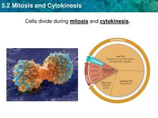
The cytokinesis block micronucleus (CBMN) assay CBMN assay was developed by Fenech and Morley in 1985 In CBMN assay Cytochalasin B, a cytoplasmic division inhibitor, is added to cell culture to identify cells that underwent one division These cells are identified as binucleate (BN) cells CBMN assay is mostly applied to peripheral blood lymphocytes (PBL), but any cell that can divide can be used CBMN assay is often used as a general toxicology test 6
The CBMN assay for radiation dose assessment Ionizing radiation is a strong clastogenic agent and thus a potent inducer of DSB and by consequence also of MN In CBMN assay for radiation dose assessment, MN are typically scored in 1000 binucleate PBL Radiation-induced MN in PBL contain mainly acentric chromosome fragments (MN-ac) resulting from non-repaired or misrepaired DNA dsb by non-homologous end joining (NHEJ) repair pathway 7
Human Lymphocytes Advantages: • Easily obtained from the peripheral blood • The majority of peripheral blood lymphocytes are in the G0 phase of the cell cycle • Phytohaemagglutinin (PHA) converts these resting lymphocytes into dividing cells by which it is possible to visualize DNA lesions in metaphase chromosomes or interphase cells after division
The MN dose response curve • Radiation-induced MN are strongly correlatedwithradiation doseandquality • The in vitro MN dose-response calibration curvesfollow the same shape as described for the standard dicentric assay: • forlow LETradiation, MN follow a linear-quadratic dose response : y = c + αD + βD2 • forhigh LETradiation alineardependence y = c + αD is observed, with high LET radiation being more effective in generating MN at the same dose levels
The MN dose response curve MN frequency /1000 cells plotted against gamma ray dose 12
MN background frequencies Drawback of the CBMN assay for applications in the low dose range: High and variable spontaneousfrequency (2-36 MN/1000 BN cells) unreliable for low dose assessment below ~ 0.2- 0.3 Gy X-or -rays 13
MN background frequencies Factors influencing the MN background levels: age, gender, diet, exposure to environmental mutagens age:systematic increase with age gender:systematically higher in females vs. males males: 0.24 - 0.44 MN/1000BN/year females: 0.52 - 0.54 MN/1000BN/year 14
MN background frequencies What do spontaneous MN contain? Analysis of MN content was performed using a FISHpan-centromeric probe the majority of spontaneous MN contain a centromere(> 70%) whole chromosome the age increaseis due to an increase in centromere-positiveMN *often the X-chromosome is involved explains the observed gender difference 15
MN background frequencies Centromere negative positive 17
Applications of the CBMN assay for biological dosimetry (1) • Chernobyl nuclear power plant and Semipalatinsk nuclear test site studies • CBMN assay was applied to assess protracted exposure, due to the incorporation of long-lived radionuclides • Conclusions:MN frequenciesmeasured in a large number of residents living in the vicinity of the nuclear sites wereincreasedandsignificantly associated with the estimated internal absorbed dose 18
Applications of the CBMN assay for biological dosimetry (2) For accidentsinvolving few people, only a limited number of MN studies are available: (1) The Istanbul accident with an unshielded former radio- therapy Co source (10 scrap metal workers) (2) Accident (Europe) with a 50 kV contact radiotherapy X ray device (one hospital worker) • Blood samples taken between 1 and 6 months after the accident Conclusions: • MN-derived dose estimates were in striking agreementwithdose values obtained from dicentricstudies 19
Applications of the CBMN assay for biological dosimetry (3) Application for biomonitoring • Several studies have been performed on large populations of nuclear power plant workers and hospital staff a dose dependent increaseof 0.0175 MN and 0.03 MN per 1000 BN cells/mSv was found in 2 different studies performed on nuclear power plant workers (receiving accumulated doses ranging from 10 to 248 mSv) Application of CBMN-centromere assay in first study resulted in increase of 0.025 MN per 1000 BN cells/mSv, with dose-dependent increasecompletely due to centromere-negative micronuclei CBMN assay, especially CBMN-centromere assay allows accumulated doses exceeding 50 mSv to be detected, at population level 20
Improvements of the CBMN assay (1) • Development of the CBMN-centromere assay to increase the low dose sensitivity • Development of the CBMN cytome assay to score more endpoints related to radiation exposure • Development of the CBMN assay for automated scoring to allow rapid analysis of large sample sizes (triage) • Development of a combined CBMN-centromere assay for automated scoring to combine high speed MN analysis with a more accurate assessment in the low dose range 21
Improvements of the CBMN assay (2) • The CBMN-centromere assay • allows discriminationbetweencentromere-positive MN(MNCM+)(whole chromosomes) andcentromere-negative MN(MNCM-)(acentric chromosome fragments) • several studies showed that the majority of spontaneous MN are CM+(70% and more), while most radiation-induced MN are CM- • the number of MNCM+ shows only a very small increase with dose • manual scoring of MNCM-lowers detection limit to 0.05 - 0.1 Gy 22
Improvements of the CBMN assay (3) • Scoring of nucleoplasmic bridges (NPBs) in the CBMN cytome assay • In CBMN cytome assay NPBs, joining two nuclei in BN cell, can be scored • NPBs originate from dicentric chromosomes 23
Improvements of the CBMN assay (4) • Advantage of scoring NPBs for biological dosimetry • NPB background frequency is lowerthan for MN and is not affected by gender • NPBs provide a direct method of measuring asymmetrical chromosome rearrangement (dicentrics, rings) in once divided cells • NPBs are more radiation specificthen MN • NPBs are increased in a dose-related mannerandcorrelatewellwith dicentric and ringchromosome frequencies analyzed in metaphase cells • analysis of NPBs is quickercompared to dicentric analysis as in the CBMN assay a large number of BN cells are accumulated 24
Improvements of the CBMN assay (5) • Automated scoring for the CBMN assay • among all cytogenetic methods, CBMN assay allows most easy and rapid scoringof radiation damage • to further increase the throughputof this technique, automated scoringprocedures have been developed • automated MN scoring is very attractive for: 1) population triage in case of large scale radiation accidents 2) for large-scale assessment of genetic damage in radiation workers receiving a high radiation burden (biomonitoring) • automated MN scoring results in a more reproducibleanalysis 25
Improvements of the CBMN assay (6) • Automated scoring for the CBMN assay • differentimage analysis systemsfor automated MN scoring are available: - MN software module integrated in the metaphase finder systemMSearch of Metasystems 1) identification of 2 adjacent similar nuclei (e.g. DAPI stained) 2) MN scoring in a circular region defined around the 2 nuclei of the BN cell - MN analysis system running on thePathFinderCellscan capture station 1) identification of the cytoplasm of Giemsa-stained cells 2) detection of the number of nuclei in the cell, allowing identification of BN cells 3) automated scoring of MN in the BN cell 26
Improvements of the CBMN assay (7) • Automated scoring for the CBMN assay 27
Improvements of the CBMN assay (8) • Automated scoring for the CBMN assay Example of BN cells with and without MN captured by the MN module for automated MN scoring of Metasystems • on 1 slide automated scoring of MN in about 2000 BN cells can be performed in less than 8 minutes Gallery of captured BN cells with MN
Improvements of the CBMN assay (9) • The CBMN assay for automated scoring • the figure shows a dose response curveobtained for automated MN scoring (by MNScore of Metasystems), based on MN data of 10 individuals • Results • dose of 1 Gycan be detected with accuracy of 0.2 Gy • 95% CI of 0 Gy and 1 Gy doses do not overlap • accurate dose estimations are also achieved at higher doses of 2 and 3 Gy Taken from Willems et al., IJRB 86(1),2-11, 2010
Improvements of the CBMN assay (10) • Development of a combined CBMN-centromere assay for automated scoring • to enhance sensitivity, reliability and processing time of MN assay by combiningautomated MN assaywithFISH pan-centromerescoring • This will allow : systematic biomonitoringof radiation workers exposed to low doses more accurate assessmentof exposure in second phaseof mass radiation casualty event - after early triage - when the time constraint will be less strict 30
Improvements of the CBMN assay (11) • Development of a combined CBMN-centromere assay for automated scoring This figure shows the average of the total number of MN (MNtotal), MNCM+ and MNCM- for 10 donors as a function of dose (up to 0.5Gy). The error bars are SEM. (modified from Baeyens et al., IJRB, 2011) 31
Improvements of the CBMN assay (12) • Development of a combined CBMN-centromere assay for automated scoring Conclusion • Semi-automated CBMN-centromere assay combineshigh speed MN analysiswith amore accurate assessmentin the low doserange, down to 0.05 Gy which makes it of special interest forlarge-scale radiation applications 32
Limitations of the CBMN assay (1) • Major limitations of CBMN assay are related to: • retrospective dosimetry • accidents involving partial body irradiation 33
Limitations of the CBMN assay (2) • Retrospective dosimetry • In some accident studies MN datawere compared with data obtained by theFISH translocationtest, which is the endpoint of choice for retrospective dosimetry • Istanbul accident (10 scrap metal workers) and accident of hospital worker with a radiotherapy X ray device (slide 19) • blood samples were taken at different time points after the accident (1month -1year) 34
Limitations of the CBMN assay (3) • Retrospective dosimetry • Results and conclusions dose estimatesobtained by scoringMN(comparable to dicentrics) arelower (20 to 30%) than dose estimates obtained by scoring translocations correctionof the MN values for the delayed blood samplingin the hospital worker (MN disappear with a half-life of 342 days) result in a dose estimatecomparableto that obtained with FISH translocations underestimationof radiation doses because MN (and dicentrics) are unstable chromosome aberrationswhich have a limited in vivo persistence 35
Limitations of the CBMN assay (4) • Accidents involving partial body irradiation • in practice, most accidents involve partial body exposures (PBE) problem with PBE: underestimationof dose because of presence ofundamaged lymphocytespresent outside the irradiation field For dicentric assay mathematical methods exist to calculate PBE • The contaminated Poisson method(Dolphin 1970) based on the finding that after whole body (WB) irradiationdicentricsarePoisson distributed • TheQdr method(Sasaki and Miyata 1968) ForCBMN assay as MN are already slightly overdispersedafter WB irradiation the applicability of a MN frequency distribution analysis with respect to a PBE is questionableand still needs further investigation 36
The CBMN assay: practical issues • Scoring criteria for the CBMN assay • Nuclear division index (NDI) • Protocols 37
Scoring criteria for the CBMN assay (1) Criteria for manual scoring of BN cells • Cytokinesis-blocked cells should have following characteristics: (a) cells should be binucleated (BN) (b) two nuclei in a BN cell should be situated within same cytoplasm (c) two nuclei in a BN cell should be approximately equal in size, staining pattern and intensity (d) two nuclei within BN cell may be unconnected or may be attached by one or more fine NPB (e) two main nuclei in BN cell may touch but ideally should not overlap each other (f) BN cells should not overlap each other 38
Scoring criteria for the CBMN assay (2) Criteria for manual scoring of MN • MN are morphologically identical to but smaller than the main nuclei. They also have the following characteristics: • diameter of MN in human lymphocytes should be smaller then 1/3rd of the mean diameter of the main nuclei • MN are non-refractile and can therefore be readily distinguished from artefacts such as staining particles • MN are not linked or connected to the main nuclei • MN may touch but not overlap the main nuclei and the micronuclear boundary should be distinguishable • MN usually have the same staining intensity as the main nuclei but occasionally staining may be more or less intense 39
Scoring criteria for the CBMN assay (3) Criteria for scoring NPB in the CBMN-cytome assay • Description is given in: - The IAEA Manual and - Cytokinesis-block micronucleus cytome assay. M. Fenech, Nature Protocols 2(5), 1084-1104, 2007 40
Nuclear division index (NDI) • In the CBMN assay, the relative frequencies of cells with 1, 2, 3, etc nuclei (NDI)can be used to define cell cycle progressionand how this is affected by radiation exposure • Calculation of the NDI NDI= M1 + M2 + M3 + M4 / N - 500 cells are scored - M1 to M4 represent the number of cells with one to four nuclei and N is the total number of viable cells scored • More details about the calculation of the NDI and its uncertainty are described in the IAEA Manual 41
Protocols (1) Standard Cytokinesis-block Micronucleus protocol • a heparinized blood sample is taken and complete culture medium (RPMI+ supplements + 10% FCS ) is prepared 42
Protocols (2) Standard Cytokinesis-block Micronucleus protocol • 0.5 ml of blood is added to complete culture medium 43
Protocols (3) Standard Cytokinesis-block Micronucleus protocol • Phytohaemagglutinin (PHA) is added as the mitogen 44
Protocols (4) Standard Cytokinesis-block Micronucleus protocol • Blood culture is incubated at 37°C and with 5%CO2 45
Protocols (5) Standard Cytokinesis-blocked Micronucleus protocol • after 24 hours incubation cytochalasin B is added to block cytokinesis 46
Protocols (6) Standard Cytokinesis-block Micronucleus protocol • Cells are harvested 3 days after culture start (between 68-72h): • Harvest protocol: • Blood cultures are centrifuged 47
Protocols (7) Standard Cytokinesis-block Micronucleus protocol • Harvest protocol: Cells are hypotonically treated with cold KCl (0.075 M) KCl 48
Protocols (8) Standard Cytokinesis-block Micronucleus protocol • Harvest protocol: After centrifugation cells are fixed with fixative solution (10 methanol / 1 acetic acid / 11 Ringer salt solution). For the automated scoring 4/1/5 ratio is used. 49
Protocols (9) Standard Cytokinesis-block Micronucleus protocol • Harvest protocol: After centrifugation cells are washed another 2 or 3 times with a fix solution containing 10 methanol / 1 acetic acid (for automation: 4/1) 50