Download
1 / 20
220 likes | 445 Views
Biology 221 Anatomy & Physiology II. TOPIC 5 Lymphatic System. Chapter 21 pp. 778-787. E. Lathrop-Davis / E. Gorski / S. Kabrhel. Functions. Transportation : Returns fluid back to blood from tissues Returns proteins back to blood from tissues

E N D
Biology 221 Anatomy & Physiology II TOPIC 5 Lymphatic System Chapter 21 pp. 778-787 E. Lathrop-Davis / E. Gorski / S. Kabrhel
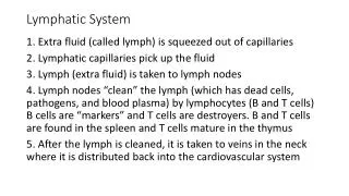
Functions • Transportation: • Returns fluid back to blood from tissues • Returns proteins back to blood from tissues • Transports fats and fat soluble vitamins (D,A,K,E) from GI tract to blood • Protection - Protects and defends body against disease (houses agranular leukocytes)
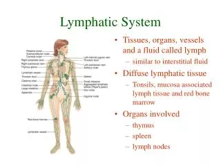
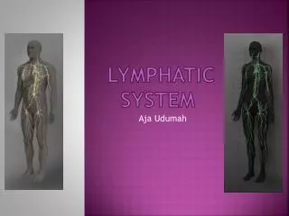
Components - Overview • Lymph – fluid • Lymphatic vessels – conduits for flow of lymph • Lymphoid tissues and associated structures - unencapsulated, less organized tissues with large numbers of white blood cells • Lymphoid organs – larger, more organized, encapsulated structures consisting largely of lymphocytes
Lymph • fluid that enters lymphatic vessels from interstitial fluid • enters lymphatic capillaries under low pressure (keeps interstitial fluid hydrostatic pressure low) • similar to blood plasma, except: • no red blood cells • more white blood cells • less protein • more fat
Lymphatic Vessels: Lymphatic Capillaries • Blind sacs consisting of simple squamous epithelium (endothelium) • Cells overlap to form valves within lumen • Cells connected by fibers to structures within tissue • Collect excess tissue fluid • Lacteals – very “leaky” lymphatic capillaries found in wall of small intestine Fig. 21.1, p. 779
Lymphatic Vessels • Lymphatics • Formed from union of lymphatic capillaries • Similar to veins, but with thinner walls, less muscle, less connective tissue, more valves • Carry lymph to lymphatic trunks • Lymphatic Trunks • Formed by union of lymphatics • Carry lymph to lymphatic ducts Fig. 21.1, p. 779 Fig. 21.2, p. 780
Lymphatic Vessels: Ducts • Formed by union of lymphatic trunks • Empty into subclavian veins on right and left • Right lymphatic duct • drains upper right quadrant of body • formed from jugular, subclavian, and bronchomediastinal trunks on right • Thoracic duct • starts as cysterna chyli (receives lymph from intestinal trunk and right and left lumbar trunks serving lower back, hips and legs) • left jugular, subclavian, and bronchomediastinal trunks add lymph from upper left quadrant of body • drains upper left quadrant, abdominopelvic cavity, legs Fig. 21.2, p. 780
Lymph Circulation • Moves along pressure gradient • Valves keep flow moving in one direction • Mechanisms believed to contribute to pressure: • “milking” by skeletal muscle (contraction of skeletal muscle puts pressure on lymphatic to move fluid forward) • pressure changes during breathing (inhalation lowers pressure in thoracic cavity, increases pressure in abdominal cavity) • pulsating of neighboring elastic arteries • contraction of smooth muscle in walls of larger lymphatic vessels and ducts
Lymphoid Tissues: Nodules • unencapsulated areas of densely packed lymphocytes in areolar connective tissue • germinal center (area of rapid cell division) surrounded by maturing cells • common in mucosae of respiratory, urinary and digestive systems http://www.usc.edu/hsc/dental/ghisto/lymp/d_75.html
Lymphoid Tissues: MALT • Mucosa-associated lymphatic tissue (MALT) • aggregates of lymphatic nodules (follicles) found in mucosae of respiratory and digestive systems • includes tonsils, Peyer’s patches http://www.usc.edu/hsc/dental/ghisto/lymp/d_69.html
Lymphoid Tissues: Tonsils • large nodules found in walls of oropharynx (lingual and palatine tonsils) and nasopharynx (adenoid or pharyngeal tonsil) http://www.usc.edu/hsc/dental/ghisto/lymp/d_44.html Fig. 23.3, p. 838
Lymphoid Organs • Larger, more organized structures consisting largely of lymphocytes • Generally encapsulated • Spleen, thymus, lymph nodes Fig. 21.5, p. 783
Lymphoid Organs: Lymph Nodes • function: filter debris, pathogens and other antigens from circulating lymph • structure: • small, ovoid, covered with connective tissue capsule • hilus • two regions: • outer cortex • inner medulla http://www.kumc.edu/instruction/medicine/anatomy/histoweb/lymphoid/lymphoid.htm http://www.usc.edu/hsc/dental/ghisto/lymp/d_1.html
Lymphoid Organs: Lymph Nodes • Cortex (C) • extensions (trabeculae; T) of capsule extend into cortex and separate it into compartments • stroma of reticular fibers supports lymphocytes • contains follicles (F) with germinal centers (GC) • Medulla (m) – contains both T cell and B cell lymphocytes and macrophages http://www.usc.edu/hsc/dental/ghisto/lymp/d_1.html
Lymph Nodes: Circulation • Afferent lymphatics – enter through capsule on cortex side • Subcapsular sinus - space between capsule and cortex filled with circulating lymph • Lymph sinuses - areas through which lymph circulates • Efferent lymphatics - leave through hilus http://www.kumc.edu/instruction/medicine/anatomy/histoweb/lymphoid/lymphoid.htm Fig. 21.4, p. 782
Lymphoid Organs: Spleen Functions: • RBC production in fetus • Stores platelets and iron • Macrophages remove abnormal and old RBCs • Helps initiate immune response to circulating antigens http://www.people.virginia.edu/~rjh9u/spleen.html http://www.kumc.edu/instruction/medicine/anatomy/histoweb/lymphoid/lymphoid.htm
Spleen: Structure • Hilus – region where blood vessels and lymphatics enter/leave • Red pulp – includes venous sinuses containing RBCs and macrophages • White pulp – contains lymphocytes (resembles lymphatic nodules) http://www.kumc.edu/instruction/medicine/anatomy/histoweb/lymphoid/lymphoid.htm Fig. 21.6, p. 784
Lymphoid Organs: Thymus • Functions: secretes hormones (thymosin and thymopoietin) that stimulate T cell lymphocytes to become immunocompetent (enables them to respond appropriately to pathogens) • most active in childhood (atrophies after adolescence) • plays a role in removing “autoreactive” lymphocytes (ones that would cause autoimmune disorders) • Location: in lower neck region (extends into mediastinum in infants)
Thymus: Structure • 2 lobes divided into lobules by extensions (septae) of the fibrous tissue capsule. Each lobule consists of: • Outer cortex of closely packed cells including dividing lymphocytes • Thymocytes (reticuloendothelial cells) • separate clusters of lymphocytes from blood (blood-thymus barrier) to keep • secrete thymic hormones (stimulate division of lymphocytes and differentiation of T cells) http://www.usc.edu/hsc/dental/ghisto/lymp/d_53.html http://www.usc.edu/hsc/dental/ghisto/lymp/d_54.html
Thymus: Structure • Inner medulla where mature T cell lymphocytes are found • T cells able to leave and enter blood or lymph • Hassall’s (thymic) corpuscles - unusual structures of unknown function http://www.usc.edu/hsc/dental/ghisto/lymp/d_59.html