Download
1 / 77
780 likes | 1.01k Views
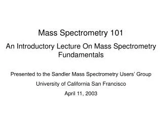
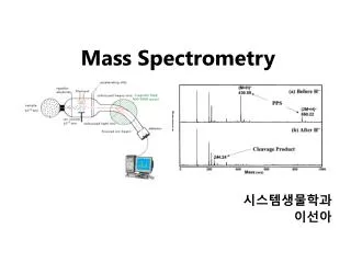
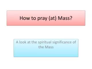
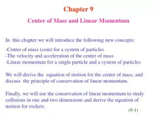
Who Needs Photons When You Have Mass?. Lecture 2. ACCA Spectroscopy Series Sept. 22, 2009 Bruce Solka , Ph.D. Sr. Principal Scientist, Unilever, retired. =. 116. 100. 90. 80. 28. 70. 88. 60. 60. 50. Relative Abundance. 46.0. 40. 30. 20. 10. 0. 80. 100. 120. 140. 160.

E N D
Who Needs Photons When You Have Mass? Lecture 2. ACCA Spectroscopy Series Sept. 22, 2009 Bruce Solka, Ph.D. Sr. Principal Scientist, Unilever, retired
= 116 100 90 80 28 70 88 60 60. 50 Relative Abundance 46.0 40 30 20 10 0 80 100 120 140 160 180 40 60 * Mass Spectrometry: ● What is it? ● Why do it? ● How does one do it? To be followed with some illustrations of practical applications *a misnomer ? (light involved only in special cases) mass
The language of mass spectrometry as used in this talk. Dalton AMU Ion Isotope Sample inlet Ion source Mass analyzer Ion trap Quadrupole Magnetic sector Turbomolecular Molecular ion Fragment ion Isotope Base peak Mean free path Electron impact Electrospray Chemical Ionization Total ion chromatogram Mass chromatogram GC-MS HPLC-MS Desorption Electrospray Collision-induced dissociation
How to provide a tour of mass spectrometry ? • Introduction • What is MS • Who am I to say • What do they look like • Who uses MS and why • How does MS work • Details of some applications Applications
1.What is mass spec? An instrument that literally measures mass of molecules Details… ● Effects of electromagnetic fields on ions made from the molecule. ● Mass of fragments of molecule also used. ●Then, the mass can be used as a tag to determine what molecules are present in a mixture and at what concentration. ASMS poster “what is mass spec?
1. What is mass spec? (cont.) • An instrument used to answer the questions of → What is present? → How much is there? Advantages over many other types of spectroscopy: “What” can range from a hydrogen atom, H mass = 1 amu), to an A,B,C transporter protein of mass 400 kD (400000 amu). “How much” can be measured in femtograms (10-15 g) attomoles (10-18 moles) and parts-per-trillion. Extraordinarily versatile, specific, and sensitive
Who am I to say what Mass Spec is? • Your Guide: Bruce Solka, Ph.D., retired • Energy (synfuels) at GTI and then to consumer products at Unilever • Postdocs in MS at U of Toronto & Purdue U • Ph.D (NIU) • Started in ca 1966…>40 years of mass spec Someday, these will be the “good old Days”
Mass Spectrometry ca. 1968 Hitachi RMU-6 Magnetic Sector Sample inlet “big” electromagnet Ion formation detector
To establish a sense of what we’ll be talking about… And because mass spectrometers come in a very wide variety of sizes and types, Next several slides are images of various types of modern mass spectrometers
Basic GC-MS: Benchtop, < $50,000 (chemist not included) to 1000 amu , most EPA work, flavors & fragrances, general organic NCIS, CSI, etc Mass Spec Gas chromatograph inlet
HPLC-MS @ $300,000Includes ESI, MALDI, APCI ionizationIdeal for proteomics studies (and most general non-volatile organic analyses). Note: Compared to last slide, MS now bigger than sample inlet
Lawrence-Livermore Labs “Accelerator Mass Spectrometer” (1MV) Isotope analysis…radiocarbon dating, geology, botany, etc. (one of 5 in U.S.)
Sensitive to isotope abundances at 1 in 1 x 10¹⁵ 10Be, 14C, 26Al, 36Cl, 41Ca, 129I http://www.physics.purdue.edu/primelab/
FTMS: Super-High resolution, mass range, and sensitivity, ($800000) Mass spec #2, FTMS, or ICR (Inside of shielded, super-con magnet} Mass spec #1… LIT-MS to form and select ions for study in… Used in: •Genomics •Proteomics •Syn. Polymers •General Organic
The Torion corp. “Guardion-7”…30 lbs and ready in 3 min. llllllII I’ll find those #@*^@ pollutants Or terrorist explosive caches, etc.
How to provide a tour of mass spectrometry ? • Introduction • What is MS • Who am I to say • What do they look like • Who uses MS and why • How does MS work • Mass analyzers • Ion sources • Details of some applications Applications
2. Why do mass spec ? • You will probably use mass spec if you’re • An anesthesiologist monitoring OR atmosphere or patient breath gases • TSA Security checking passengers for explosive residues. • A biochemist determining physical location of a drug metabolite within a cell. • A Crime lab analyst identifying a greasy residue. • Geochemist determining the age of a petroleum reservoir. • Archeologist identifying a resin in an artifact. • Art historian determining a pigment in authentication of a painting. • Chemist determining the cause of a malodor in a consumer complaint. • Chemist determining why a blue shampoo has turned green. • Molecular biologist determining amino acid sequence of a new protein. • USA PFC responsible for detecting presence of any nerve gas on the battlefield. • Environmental analyst determining whether a cancer cluster may be due to carcinogens in the air, water, or soil. Mass spec, if practiced in all of it’s various modes is extremely versatile!!! • Is there really water on Mars?
Project Phoenix Mars Lander (2008) Magnetic sector mass analyzer Sample ovens and ion source Detector
How to provide a tour of mass spectrometry ? • Introduction • What is MS • Who am I to say • What do they look like • Who uses MS and why • How does MS work • Mass analyzers • Ion sources • Details of some applications
2. How does it work? 2.1. Mass Analyzers mass range?, mass resolution? Sensitivity? • Magnetic sector MS • Quadrupole MS • Ion trap MS • Quadrupole • Linear • Time of Flight MS • FTMS (Ion cyclotron resonance) • Orbitrap™ • Hybrids of above ALL depend on mass-dependant interaction of ions with electromagnetic fields.
eV = ½ mv2 m/q = R2B2/2v Sample inlet Ion Source mass analyzer detector Data aq. Mean free path Mean free path, L = 1/nσ n is number of molecules per unit volume σ, the collision cross-section, relates to size http://www.cem.msu.edu/~reusch/VirtualText/Spectrpy/MassSpec/masspec1.htm
The Turbomolecular Vacuum Pump Turbine blades, 65,000 rpmyou Attaches to MS flight tube ca 1 x 10⁻⁷torr 1 atm = 760 torr 1 x 10⁻⁷torr = 76 millionths of atmosphere To “rough” vacuum pump (ca 0.01 torr) When Ions fly, you generally don’t want them hitting gas molecules
Quadrupole Mass Analyzer http://www.chem.vt.edu/chem-ed/ms/quadrupo.html
Ion Trap mass analyzer Sample Inlet Ion source “shutter electrode” End cap • Fill trap • Eject all but target ion • Use fields to increase target ions energy until it fragments • Ramp end caps to get spectra of target fragment • Continue Ring electrode • Fill trap • Ramp end cap voltage • Ions exit to detector in order of mass • Refill trap for next scan + + + Detector RF Mass Spectrum MSn
Sequential Mass Spectrometry in the Ion Trap MS (glycolipids are carbohydrate/lipid conjugates that occur in cell membranes) From “Ion Trap Mass Spectrometry” Wong and Cooks, http://www.currentseparations.com/issues/16-3/cs16-3c.pdf
Again, ALL Mass spectrometers must have: • sample inlet (atmospheric to high Vac.) • Ion source • mass analyzer • detector • data acquisition (and inst. Control and library searching ) Next, we’ll look at the three most common ion sources • Electron impact • Chemical Ionization • Electrospray ionization
3. How does MS work? 3. B. Ion Sources • Electron Impact (EI) • Chemical Ionization (CI) • Electrospray Ionization (ESI) • Atmospheric Chemical Ionization (APCI) • Matrix-assisted Laser Desorption (MALDI) • Inductively coupled plasma Discharge (ICP) • Desorption Electrospray Ionization (DESI) (Plus a half-dozen more that are no longer commonly used). No.s 3-7 also double as sample inlets
M M+ (5 amps) M M + M = 116 100 90 80 28 70 88 60 60. F1+ + N1 F2+ + N2 Etc. 50 Relative Abundance 46.0 40 M+* 30 20 10 0 80 100 120 140 160 180 40 60 mass 1. Electron impact ionization e- + M M+ + 2 e- (ionization potential 9 – 20 eV} M+ filament To mass analyzer V 10 to 100 V Excess energy in molecule-ion? Isotope peaks “Fragmentation”, giving the mass spectrum of the molecule.
Isotopes, Molecular ions, and Fragments 75.8% of natural Chlorine atoms are atomic weight 35 25.4% are atomic weight 37. (vinyl chloride) Mass Spectrum shows: • Two molecular ions in 75.8 / 25.4 ratio. • Chlorine-containing fragments in same ratio (note m/z 47/49). • Hydrocarbon fragments clearly lack this (27/29? Nope) http://www.cem.msu.edu/~reusch/VirtualText/Spectrpy/MassSpec/masspec1.htm
True Peak Shape on Quadrupole MS (Ben) Pkwdth_080919110220 # 67 RT: 1.12 AV: 1 NL: 9.77E6 T: {0,0} + p EI det=302.00 Full ms [ 15.00-300.00] x50 147.3 100 90 80 70 Relative Abundance 60 Isotope peaks M+1, m+2,etc. 50 40 150.3 30 20 148.3 151.3 10 149.2 152.3 153.2 154.2 146 147 148 149 150 151 152 153 154 m/z
It all comes from thermodynamics and kinetics Stable CO2 molecular ion dominates spectra Frags present but small. Lots of fragments in these but quite different spectra for similar compounds.
Interpretation of EI mass spectra Before computers…. • Many long hours spent correlating spectra to molecular structure to develop predictive correlations. • Hero’s spent their lives recording spectra of thousands of compounds relevant to their work. • Positive id of an analyte required comparison with spectra of authentic standard on same MS at same time. Since about 1975…. Computer-searchable libraries of mass spectra …225,000 entries Search results list 10 or 25 most probable compounds. Chromatographic inlets add retention time information. If you’re going to court, positive id of an analyte still requires comparison with spectra of authentic standard.
Library Search Output Screen photo…software dosen’t permit copying to other files If not the actual compound, probably chemically related
2. Chemical Ionization, a gentle ionization • Formation of reactant ion (methane, for example). • (E.I. at higher pressure of methane….ca 1 torr) • CH4+ + CH4 CH5+ + CH3. • C2H5+ + H2 + H+ • Reaction with sample molecule. • CH5+ + M MH+ + CH4 ∆E • Excess energy now limited by ∆E • • less excess energy in MH+ • • spectrum simplified, often to MH+ only. This is shown mainly as an example of the fact that various types of reactive chemistry can be studied or used in the ion source!!
Electrospray Ionization: • an even more gentle technique. → Perhaps most important development in MS ever (MALDI?). → Expanded upper mass limit to 100s of thousands for peptide/protein and synthetic polymer work. → Permitted study of protein structure/function in new field of proteomics. → Also useful for many other non-volatile compounds in the 1000 – 4000 AMU M. Wt. range (lipids, triglycerides,.etc.) Biochemical and polymer applications of ESI first developed by Prof. John Fenn, Yale U. 2002
3. Electrospray Ionization, continued Mass spec ion optics 250000 0.001 to 2 ml/min liquid flow (e.g. from HPLC) 200000 Intensity 150000 + 4000eV 100000 ++ ++ ++ ++ ++ + + + + + Vac. Pump 10-2 torr Vac. Pump 10-5 torr ++ ++ ++ ++ + + + + + + + + + ++ ++ ++ ++ + + + + + + + + 50000 + + + + + + + + + + ++ ++ ++ ++ ++ ++ ++ 50 100 150 200 250 300 350 400 450 500 5 µ? mass 2. Droplet stabilizes charge by distorting 1. Aerosol evaporation builds charge density 50 µ + + + + BTW… the aerosol tip Metal syringe needle (for gas) Very gentle, only molecular ions in spectrum 3. Droplet ejects ions by coulombic explosion Silica capillary for sample solution (HPLC?)
Sequencing of peptides separated by 2D-gel electrophoresis of digests “creature” material Credit: Zina Deretsky, National Science Foundation http://www.thermo.com/com/cda/resources/resources_detail/1,2166,200553,00.html
Sample Needed for ESI-MSn (This is a comfortable amount for service work…not to be confused with detection limits) • Dry Sample* (preferred) In Solution Protein/Peptides 1µg 1µg/100µL (0.001% or 10 ppm) • Polymers 1 mg 1mg/250µL ( 0.4%) • Dendrimers 1 mg 1mg/250µL • Organometallics 1 mg 1mg/250µL • Organics 1 mg 1mg/250µL • For quantitative measures, • enough to weigh is more than enough for MS These figures are more than enough for other types of MS Sensitivity such as this is one of the principal advantages of MS over other chemical analysis techniques.
ESI mass spectrum of horse heart myoglobin (mass 16955 Da) So why does a 16,955 Da protein give a spectrum with these low masses? The key is that all mass spec interactions are on the basis of m/q ratio (16955 Da + 12 protons) / 12 charges = mass 1413.9 (16955 Da + 13 protons) / 13 charges = mass 1305.1 (16955 Da + 14 protons) / 14 charges = mass 1212.1 etc. Note general accuracy for weighing this molecule goes to 5 sig. figs. www.chm.bris.ac.uk/ms/theory/esi-ionisation.html
How Does MS Work - Summary 1. All MS must have combination of these key components: 2. Table only includes most common types…there are many others out there.
How to provide a tour of mass spectrometry ? • Introduction • What is MS • Who am I to say • What do they look like • Who uses MS and why • How does MS work • Mass analyzers • Ion sources • Details of some applications
Details of some applications • Mass chromatograms used to find source of odor in pkg. material. • LC-MS to determine reason for “oily” antiperspirant package. • GC-MS to find cause of malodorous hair spray. • ESI-IT-MS2 to determine structural details of a cationic surfactant. • How LC-ESI-IT-MS2 is used in protein sequencing.
Example 1: Source of problem odor found by “mass chromatograms”. Inlet: GC-MS MS: Quadrupole Ionization: EI Approach: Gas headspace analysis at sub-ppm concentrations Sensitivity Required: Human nose sensitive to parts-per-billion of some compounds A 1 cc sample of air weighs about 10-3 grams A Quad GC-MS in good shape can detect picograms (10-12 grams) 10-12 grams compound X = 10-9 grams X per gram sample 10-3 grams sample This is one part per billion
Odor is molecular Something smells funny here Human nose: ppb At ppb level, Hundreds of compounds http://www.cf.ac.uk/biosi/staff/jacob/teaching/sensory/olfact1.html
Solid Phase Microextraction(SPME) For concentration of sub-ppm organic analytes in air or water Silica fiber Plunger Absorbant coating such as GC stationary phase
SPME sampling of roll-on headspace? Malodor found to be due to polypropylene applicator ball by olfactory testing (i.e., they stunk and we had 200,000 of them)
Roll-on AP Applicators are hollow polypropylene spheres Volatiles from polypropylene will tend to concentrate within sphere
GC-MS Inlet Configuration Gas Chromatograph Mass spectrometer time
NL: 17.38 12 ball6 10 “Good” 19.22 17.14 8 15.50 Relative Abundance 14.76 6 18.59 15.67 4 15.88 14.09 12.47 2 0 NL: 14.73 15.47 15.70 17.32 18.84 1.78E7 16.84 12 TIC MS Major differences ball4 10 “Bad” 19.81 8 14.07 6 Minor, but interesting difference. 18.37 4 12.61 13.59 2 0 12 13 14 15 16 17 18 19 Time (min) GC-MS (EI) Headspace Comparison of Polypropylene Balls 4.80E7 TIC MS Remember…every second on chromatogram contains a complete mass spectrum at that time. These are “total ionization chromatograms” (TIC)