Download
1 / 29
300 likes | 641 Views
Biosynthesis and degradation of proteins. Bruno Sopko. Content. Proteosynt hesis Post-translation processing of proteins Protein degradation. Proteosynthesis. Aminoacyl-tRNA formation Iniciation Elongation Termination. Aminoacyl - tRNA formation.

E N D
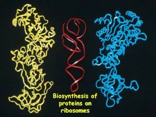
Biosynthesis and degradation of proteins Bruno Sopko
Content • Proteosynthesis • Post-translation processing of proteins • Protein degradation
Proteosynthesis • Aminoacyl-tRNA formation • Iniciation • Elongation • Termination
Aminoacyl-tRNAformation Amino acid + ATP ↔ Aminoacyl-AMP + PPi Aminoacyl-AMP + tRNA ↔ Aminoacyl-tRNA + AMP • Each amino acid has „its own“ tRNA and aminoacyl-tRNAsynthetase (ARS) • Reactions are cytosolic • Errors are corrected by specific correcting enzymes • ARS have other enzyme activity (other signaling molecule?)
Post-translationprocessingofproteins • Secondary structure and role of the chaperons • Proteolytic modifications • Glycosylation • Other modifications (hydroxylation, phosphorylation, acetylation, methylation, carboxylation)
Transmembrane proteins - examples • Type I – glycophorin, LDL receptor, influenza HA protein, insulin receptor, growth hormone receptor … • Type II – transferrin receptor, influenza HN protein, Golgi sialyltransferase, Golgi galactosyltranferase … • Type III – cytochrome P450 … • Type IV – G-protein, glucose receptors (GLUT 1 …), connexin, voltage gated Ca2+ channel …
Protein disulfidisomerase (PDI) and peptidylprolylcis-izomerase PPI:
Other modifications • O-glycosylation • Hydroxylation (hydroxyproline, hydroxylysine) • Methylation (mono- , di- and even trimethyllysine) • PHOSPHORYLATION • Carboxylation (γ-carboxyglutamate, vitamin K, fibrinogen) • Acetylation • ……..
Protein degradation • Proteases • Protein degradationsystems • Ubiquitin and proteasome • Activation of proteases • Proteaseinhibitors
Proteases • Serine proteases(trypsin, chymotrypsin, elastase ….) • Aspartateproteases(pepsin, some proteases found in lysosomes, renin, HIV-protease …) • Metalloproteases(carboxypeptidases, various matrix metalloproteases …) • Cysteine proteases(papain, cathepsins, caspases, calpains …)
Protein degradationsystems • Vacuolar (lysosomes, endosomes, ER, …) • Ubiquitin pathway (proteasome)
Activationofproteases • Most proteases are synthesized as larger pre-proteins. During activation, the pre-protein is cleaved to remove an inhibitory segment. • In some cases activation involves dissociation of an inhibitory protein • Activation may occur after a protease is delivered to a particular compartment within a cell or to the extracellular milieu. • Caspases involved in initiation of apoptosis are activated by interaction with large complexes of scaffolding and activating proteins called apoptosomes.
Proteaseinhibitors • IAPs are proteins that block apoptosis by binding to and inhibiting caspases. The apoptosis-stimulating protein Smac antagonizes the effect of IAPs on caspases. • TIMPs are inhibitors of metalloproteases that are secreted by cells. A domain of the inhibitor protein interacts with the catalytic Zn2+. • Cystatins are inhibitors of lysosomalcathepsins. Some of these (also called stefins) are found in the cytosol and others in the extracellular space. Cystatins protect cells against cathepsins that may escape from lysosomes. • Serpins are widely distributed proteins that utilize a unique suicidemechanism to inhibit serine or cysteine proteases. A large conformational change in the serpin accompanies cleavage of its substrate loop. This leads to disordering of the protease active site, preventing completion of the reaction. The serpin remains covalently linked to the protease as an acyl-enzyme intermediate. • Non-specific: α2-macroglobulin
Literature • Marks´ Basic Medical Biochemistry, A Clinical Approach, third edition, 2009 (M. Lieberman, A.D. Marks) • B. Wilkinson, H.F. Gilbert / Biochimica et BiophysicaActa 1699 (2004) 35–44 • F. Ulrich Hartl, Andreas Bracher & ManajitHayer-Hartl, Molecular chaperones in protein folding and proteostasis, Nature 475 (2011)