Download
1 / 46
460 likes | 480 Views
Explore different methods of cell mutant selection, including auxotrophs, drug resistance, antibodies, visual inspection, FACS, and temperature-sensitive mutants. Learn about the use of cell fusion and heterokaryons for gene mapping and regulation. Complementation analysis and mapping genes to chromosomes are also discussed.

E N D
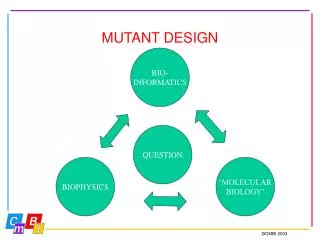
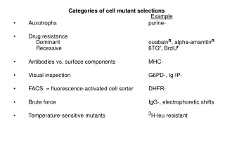
Categories of cell mutant selections Example • Auxotrophs purine- • Drug resistance Dominant ouabainR, alpha-amanitinR Recessive 6TGr, BrdUr • Antibodies vs. surface components MHC- • Visual inspection G6PD-, Ig IP- • FACS = fluorescence‑activated cell sorter DHFR- • Brute force IgG-, electrophoretic shifts • Temperature‑sensitive mutants 3H-leu resistant
Purine biosynthesis, salvage pathways, and inhibitors Adenine(A) (diaminopurine) (8-azaadenine) Methotrexate Folate (=amethopterin) (~aminopterin) Adenosine APRT Adenosine FH4 kinase Nuc. Acid AMP Glycine Thymidine (T) Adenylosucc. Alanosine PRPP + IMP glutamine HGPRT Azaserine XMP GMP Nuc. Acid Hypoxanthine Glutamine XGPRT HGPRT (H) (Eco gpt) Guanine Xanthine Mycophenolic (6-thioguanine) (X) acid (8-azaguanine) Salvage enzymes Biosynthesis; Analogs (iytal.) Code: Inhibitors (drugs, in italics) PRPP = phosphoribosyl pyrophosphate; FH4=tetrahydrofolate
Purine biosynthesis, salvage pathways, and inhibitors Adenine(A) (diaminopurine, DAP) (8-azaadenine, 8AA) Methotrexate Folate (=amethopterin) (~aminopterin) Adenosine APRT Adenosine FH4 kinase Nuc. Acid AMP Glycine Thymidine (T) Adenylosucc. Alanosine PRPP + IMP glutamine HGPRT Azaserine XMP GMP Nuc. Acid Hypoxanthine Glutamine XGPRT HGPRT (H) (Eco gpt) Guanine Xanthine Mycophenolic (6-thioguanine, 6TG) (X) (8-azaguanine, 8AG) Test yourself: Fill in the boxes Grow (+) or not grow(-) Click for the answers col. by col. Growth pattern examples GHT = glycine, hypoxanthine, and thymidine A = adenine H = hypoxanthine G = glycine TG = 6-thioguanine (G analog) DAP = diaminopurine (A analog) MTX = methotrexate (DHFR inhibitor) DHFR = dihydrofolate reductase HPRT = hypoxanthine-guanine phosphoribosyltransferase APRT = adenine phosphoribosyltransferase in italics + + - - - + - + - - - + + - + + + + - + + - - - - + - - - + + -
Cell mutant types: • 1. Auxotrophs (BrdU reverse selection, not discussed) • 2. Drug resistance (dominants or recessives) • 3. Temperature‑sensitive mutants: cell cycle mutants. • Tritiated amino acid suicide (aa‑tRNA synthetases) • 4. Antibodies. Lysis with complement. Targets cell surface constituents mostly (e.g., MHC) • 5. Visual inspection at colony level: • A. Sib selection (G6PD) • B. Replica plating (LDH) • C. Secreted product (Ig: anti-Ig IP) • FACS = fluorescence‑activated cell sorter (cell surface antigen or internal ligand binding protein) • Brute force (clonal biochemical analysis, e.g., electrophoretic variants (e.g., Ig, isozymes)) • MHC = major histocompatability locus or proteins • G6PD = glucose-6-phosphate dehydrogenase; • LCH = lactate dehydrogenase; Ig = immunoglobulin. IP = immunoprecipitate
Cell fusion (for gene juxtaposition, mapping, protein trafficking, etc. ) Fusogenic agents PEG, Sendai virus (syncytia promoting, as HIV). Heterokaryons (2 nuclei), no cell reproduction (limited duration). (e.g., studied membrane fluidity, nuclear shuttling, gene activation (myoblasts) Hybrids (nuclei fuse, some cells (minority) survive and reproduce). Small % of heterokaryons. Complementation (e.g., auxotrophs with same requirement) allows selection Dominance vs. recessiveness can be tested. Chromosome loss from hybrids Mapping: chromosome assignment. Synteny. Radiation hybrids: linkage analysis (sub-chromosomal regional assignments). PEG =polyethylene glycol, (available 1000 to 6000 MW)
Cell fusion Hprt+, TK- + Parental cells Hprt-, TK+ HAT- HAT- PEG (polyethylene glycol, mw ~ 6000 Sendai virus, inactivated Cell fusion Heterokaryon (or, alternatively, homokaryon) HAT medium Hprt-, TK+ Hprt+ TK- Hybrid cell HAT+ Cell cycle, Nuclear fusion, Mitosis, Survival, reproducton Hprt-, TK+, Hprt+ TK- Heterokaryon use examples: membrane dynamics (lateral diffusion of membrane proteins) shuttling proteins (e.g., hnRNP A1 ), gene regulation (e.g., turn on myogenesis) • Hybrid cells: examples of use: • gene mapping (synteny) • gene regulation (extinction) • complementation (pyrimidine path) Synteny = genes physically linked on the same chromosome are syntenic.
Frye and Edidin, 1970: Use of cell fusion and heterokaryons to measure the difusio of membrane proteins Complete mixing in < 40 min. No diffusion at low temperature (<15-20 deg) http://www.erin.utoronto.ca/~w3bio315/lecture2.htm
Complementation analysis Mutant parent 1 Mutant parent 1 Mutant parent 2 Mutant parent 2 gly2- + + gly3- gly1- gly1- Cell fusion Cell fusion Hybrid cell Hybrid cell glyA- glyB+/glyA- glyB+ glyA- glyB+-/glyA+ glyB- Glycine-free medium: No growth No complementation Same “complementation group” same gene (named glyA) Gly1 and gly2 are 2 alleles of gene glyA Glycine-free medium: Yes, growth Yes, complementation different genes genes (named glyA and glyB) Gly1 is in gene glyA Gly3 is in gene glyB
Mapping genes to chromosomes (hybrids) Hprt- x tk-Hybrid cell (Human x Rodent) Reduced hybrid missing some human chromosomes Spontaneous chromosome loss (human ~ preferentially lost) Hprt-TK+ / Hprt+TK- Just passage and wait Hprt-TK+ /Hprt+TK- Correlate identified chromosome loss ( ) with loss of phenotypic trait (isozyme, DNA sequence, etc.) Isozymes = enzyme variants that can be distinguished from each other by physical properties, often electrophoretic mobility in native gels (net charge).
Radiation hybrids Ionizing radiation fragments the human donor cell chromosomes After fusion, some fragments are integrated into the rodent chromosomes. Checking these “reduced” hybrids for human markers (DNA restriction fragments, PCR products, or isozymes) allows conclusion about genetic linkage, the more often two markers are integrated together the closer the linkage., x Select for a human gene (e.g., hprt) to eliminate rodent parental cells (e.g., x= hprt-) Irradiated human cells die
Ted Puck: mutagenesis; auxotrophic mutants in CHO cells (U. Colo.) Mary Weiss:turning off differentiation genes in cell hybrids (Institut Pasteur) Helen Blau: turning on muscle genes in heterokaryons (Stanford) Michael Edidin: 2-D diffusion of proteins in the cell membranein heterokaryons (Johns Hopkins) Frank Ruddle: mapping by chromosome segregation from cell hybrids.(Yale) nuclear-cytoplasmic shuttling in heterokaryons (Penn)
DNA transfection Transfection agents: CaPO4 (co-precipitates with DNA) Electroporation (naked DNA, high voltage pulse transient holes) Lipofection (multilamellar liposomes) Polybrene (cationic detergent) Ballistic (DNA-coated gold particles) DEAE-dextran (toxic, OK for transient) Poly-ethylenimine (cheap) Must traverse cytoplasm. Much engulfed in lysosomes. Inhibition of lysosomal function often helps (chloroquin). Co-integration of high MW DNA . Can = 2000 KB. Separate plasmids transfected together same site (co-integration). Separate transfections separate locations Random or semi‑random (many) integration sites (unless targeted) Low but real homologous recombination rate. History: mammalian cell transfection developed for practical use at Columbia (at P&S: Wigler, Axel and Silverstein) DEAE= diethyl-amino-ethyl (positively charged); dextran =~ poly-glucose
Mike Wigler Richard Axel Saul Silverstein History: discovered for practical use at Columbia (P&S: Wigler Axel and Silverstein)
Viral transduction of genes Lentivirus (a retrovirus) env gag pol RNA +strand genomes (2) Also, adenovirus (DNA)
DNA Packaging signal on RNA Viral structural proteins contributed by packaging cell line (gag, env, pol, etc.) Reverse transcribes to cDNA, integrates Infects cells at high efficiency http://en.dogeno.us/2009/11/concentrate-retrovirus-carrying-vsv-g-envelope-by-ultracentrifugation/
Transient transfection vs. permanent: cloned genes Unintegrated DNA chromosomally integrated. Unnatural? Position effects ? Super-physiological expression (so analyze a pool of many to levels (per transfected cell) ? average) Transient -> 10‑90% transfection efficiency (stain) Permanents more like 0.001 transfectants per μg DNA per cell (~high). i.e., 106 -> 1000 colonies; could be much less for certain types of cells
One the most dramatic first applications of gene transfection from total DNA: Transfer of the growth‑transformed phenotype: ability to grow in multilayers or in suspension in soft agar (Weinberg, Wigler): DNA from tumor transfected into growth-controlled mouse 3T3 or Rat1 cells. Look for foci (one = focus). Make a genomic library from growth‑transformed transfectant. Screen for human Alu repeat by colony hybridization. Verify that cloned DNA yields high frequency of focus‑forming transfectants. Isolate cDNA by hybridization to the cloned genomic DNA. Sequence. Identify gene: = a dominant oncogene. Ras, a signaling protein in a transducing pathway for sensing growth factors Transformed Mouse 3T3 cells transfected with an EGFreceptor gene Mouse 3T3 cells
Recombination; gene targeting Mitotic recombination between homologous chromosomes; relation to cancer through the loss of tumor suppressor genes LOH = loss of homozygosity: WT = +/+ mutation +/- (WT phenotype) (LOH via homologous recombination in G2; or chromosome loss and duplication) -/- (mutant phenotype revealed) Recombination of transfecting genes: homologous (rare) vs. non‑homologous (common) recombination.
Gene knockouts via homologous recombination Resistant to gancyclovir ES cells and transgenic mice. Selection for homologous recombinants via the loss of HSV TK genes (Capecchi): –tk – homol. region – YFG – homol. region – tk – (YFG = your favorite gene) Gene X in figure at right. Non-homologous recombination favors ends; tk is inserted, conferring sensitivity to the drug gancyclovir (HSVtk specific, not a substrate for human tk) Die in gancyclovir HSV-TK gene is removed during homologous recombination, but remains joined during non-homologous recombination. Unlike mammalian TK, HSVTk converts gancyclovir to a toxic product HSV = Herpes simplex virus; tk = thymidine kinase; FIAU = equivalent to gancyclovir, today M. Capecchi, Nature Medicine7, 1086 - 1090 (2001) Generating mice with targeted mutations
Most work in ES cells mice homozygosis via F1 breeding Little work in cultured lines: Allele replacements in cultured cell lines (e.g., APRT). Myc double sequential K.O. = viable, ~sick (J. Sedivy) Splicing factor (ASF) double K.O. see next graphic. APRT = adenine phosphoribosyltransferase ASF = alternative splcing factor
neo One ASF gene allele disrupted by homologous recombination ASF ASF neo hol ASF- hol ASF ASF ASF Human Human Human Human Human pur ASF ASF ASF ASF ASF Both alleles have been disrupted in some neoR, holR cells neo ASF- neo +tet pur ASF- pur X Cell dies without ASF(follow events biochemically –surprise = genomic instability) cell viable(covered by human ASF gene) Double knockout of the ASF gene, a vital gene, by homologous recombination Chicken DT40 cells + neoZ neo Tet-off promoter Hol = histidinol resistance; pur = puromycin resistance Drug resistance genes here chosen for illustration. Wang, Takagaki, and Manley, Targeted disruption of an essential vertebrate gene: ASF/SF2 is required for cell viability. Genes Dev. 1996, 10:2588-99.
NAD+ Histidinol dehydrogenase Histidinol dehydrogenase detoxifies histidinol, confers histidinol resistance protein synthesis inhibits protein synthesis (competes with histidine)
Methotrexate (MTX, amethopterin) Gene amplification for high level production in CHO dhfr- cells. DHFR system (dihydrofolate reductase): Selection for resistance to marginal levels of methotrexate DHFR DHFR Folate tetrahydrofolate dihydrofolate Glycine Purine nucleotides (AMP and GMP) Thymidylic acid (TMP) MDR “FH4” “FH2” Resistance to MTX can occur via 3 different mechanaisms: 1) Methotrexate permeation mutants (incl. MDR, increased efflux)) 2) Altered DHFR with lower MTX binding affinity 3) Overproduction of DHFR protein MDR = multiple drug resistance
Gene amplification: dhfr • Historically: • Methotrexate resistance • MTX inhibits dihydrofolate reductase (DHFR) • MTX-resistant cells have (in order of discovery, 1970’s): • High DHFR enzyme activity • High DHFR protein • High protein synthetic rate • High translatable mRNA • High mRNA level (by hybridization) • High DNA level. • Homogeneously staining, expanded chromosomal regions (HSRs) • HSRs are the location of the high number of dhfr genes. • Double minute chromosomes are an occasional alternative form. • Amplicons (distance between repeated genes) are large (300 KB). • (dhfr gene = ~ 25 kb) • HSRs can shrink, migrate.
Tritium grains from hybridized cDNA Gene amplification HSR: Homogenously staining region Nunberg et al. PNAS 1980 (Schimke, Sci. Amer.)
Gene amplification “Homogeneously staining region” FISH, here FISH = fluorescent in situ hybridization
+FISH Original locus? HSR dmin upon DS break induced by a homing endonuclease (I-SceI). Restriction-type enzyme with a very long recognition sequence ( ~20 bp) HSR = homogeneously staining region Dmin = double minute chromosomes Arnaud Coquelle, Lorène Rozier, Bernard Dutrillaux and Michelle Debatisse ONCOGENE, 31 October 2002, Volume 21, Number 50, Pages 7671-7679 Induction of multiple double-strand breaks within an hsr by meganuclease I-SceI expression or fragile site activation leads to formation of double minutes and other chromosomal rearrangements
Ampification models: over-replication, unequal sister chromatid exchange, breakage and fusion (Tanaka et al.). Map dhfr amplicons (Schimke, Hamlin): ~ 300 kb , but wide range Gene amplification is rare in normal cells (Wahl, Tslty). p53- mutation allows it. In nature:rDNA in oocytes; Drosophila chorion genes. In medicine:chemotherapy resistance (MDR, P-glycoprotein, efflux pump) cancer (myc, ras) In biotechnology:high level recombinant protein production in mammalian cells MDR = multiple drug resistance
Joyce Hamlin Bob Schimke John Littlefield Some notable gene amplification players Fred Alt Geoff Wahl George Stark
Gene amplification for high level recombinant protein production in mammalian cells. Principal system = dhfr- CHO cells Facilitated by the availability of DHFR-deficient mutant CHO cells CHO dhfr- cells + vector with dhfr minigene + YFG -GHT medium Most cells die. Transfectants live. + gradually increasing concentrations of MTX Cells with gradually amplified dhfr transgenes survive. YFG is co-amplified along with the dhfr minigene. -GHT = without glycine, hypoxanthine (a purein source) and thymdine
DHFR- cells require G,H,T 3H-DNA X 3H-dUMP and are resistant to tritiated deoxyuridine X X DHFR- cells selected by their resistance to radioactive 3H-deoxyuridine: 3HdU 3HdUMP 3H-TMP 3H DNA death from radioactive decay. DHFR- cells require glycine, hypoxanthine and thymidine (GHT). In GHT-free medium CHO dhfr- cells die, but transfectants that have received a dhfr minigene, +/- YFG, survive.
A different major system for high level Mab production NS0 cells: Mouse myeloma cells, high IgG producers IgG- variants = NS0 No endogenous IgG, but cell is a natural IgG secretor. Lack glutamine synthetase (GS): glutamate + NH3 + ATP glutamine + ADP + Pi Vector = MAb genes driven by strong promoters (H-chain, L-chain) + GS cDNA gene (Bebbington) Select on glutamine-free medium Inhibit GS with methionine sulfoximine (gln analog) Select for GS overproducers --->--> (gene amplification does not seem to be operating in this system of the GS cDNA gene and linked Mab genes) Proprietary (Lonza Biologics)
Transfection strategies • YFG (Your Favorite Gene) linked to a dhfr minigene on a single plasmid A. ~Insures co-integration B. ~Insures co-amplification • YFG and dhfr on separate plasmids A. Allows a high ratio of YFG to dhfr to start B. Co-amplification not assured but commonly occurs.
Linked amp CHO cells
Co-amp3 (with or without pre-ligation)
kaufman Y.F.G. DHFR Dicistronic mRNA (ribosome read-through) Also, later, better dhfr translation using an IRES, Internal ribosome intiation site, used mostly in viral but also in some cellular genes. In theory, not an advantage. Y.F.G. DHFR IRES
Amplification protocol Note: Process is lengthy and tedious.
Some marketed recombinant proteins Erythropoietin (Epogen, Procrit) J&J, Amgen Tissue plasminogen activator (TPA) Genentech Growth Hormone (Genentech) Insulin (Genentech) Beta-interferon (Avonex) Biogen-IDEC Alpha-interferon (IntronA) Schering-Plough Neupogen (Amgen) Etanercept – TNF receptor + IgG (Enbrel) Amgen Monoclonal antibodies (mAbs): see next