CASE 2
CASE 2. Sharon, a 32-year old woman, presents to you, her GP with earache. Whilst examining her ear you notice a lump in her neck that moves when she swallows. You think that she might have a thyroid nodule. .

CASE 2
E N D
Presentation Transcript
Sharon, a 32-year old woman, presents to you, her GP with earache. Whilst examining her ear you notice a lump in her neck that moves when she swallows. You think that she might have a thyroid nodule. Q1 Assuming that you are correct, what specific questions would you like to ask Sharon?
Want to distinguish between benign & malignant • (Full Hx as normal) • Ever noticed it before? If so for how long? • Has it grown? If so, over what timeframe? • Painful? Describe. • Dysphagia? Odynophagia? Hoarse voice? • Hx radiation? FHx of thyroid cancer? • Sx of hyperthyroidism (wt loss, anxiety, sweating etc.) – Malignant usually non-functioning
Sharon says that until you pointed it out, she had been unaware of any swelling in her neck. She says that she has been anxious and stressed for some time, but she attributes this to having three young children and studying for a PhD. Otherwise there is nothing else of note in her history. You then turn to examine Sharon. • Q2 What features would you look for when examining a patient with a lump in the neck like Sharon?
General inspection • Neck specific inspection • Position, generalised or localised swelling, pulsations, movement on swallowing, erythema etc. • Palpate (now assuming its thyroid) • Size, shape, consistency, mobility, tenderness. • Thrills, bruits, percussion, enlarged LN’s • You know all the rest!
On examination, Sharon is clinically euthyroid. Neck palpation reveals a soft, 2cm mobile nodule in the right lobe of thyroid gland with no fixation to adjacent tissues and no palpable lymph nodes.You arrange for Sharon to have her TSH levels checked. The test results come back the next day and demonstrate a TSH that falls within the normal range (1.8 mU/L [0.40-5.00mIU/L]) • Q3 On the basis of these results what would be your next line of investigation and why?
Investigating the lump… • FNAC is first line • U/S
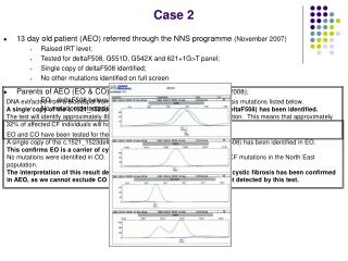
Q4 How would you have proceeded if Sharon had been found to be thyrotoxic? What information might be provided by this line of investigation? • Need to distinguish between a toxic (hot) nodule causing thyrotoxicosis - unlikely to be malignant • OR an overactive thyroid (i.e. Grave’s) with a cold nodule – cold nodules more likely to be malignant in a pt with Graves • To distinguish: patient undergoes a thyroid uptake scan (RAIU) – pt swallows radioactive iodine, and its absorption by the thyroid is studied 6 and 24hrs later. • Diffuse bilateral uptake = Grave’s dz • Irregular uptake = toxic multinodular goitre • Diminished uptake = thyroiditis • Solitary focal uptake = toxic adenoma
A = normal • B = Grave’s • C = toxic multinodular goitre • D = toxic
An ultrasound of Sharon's thyroid gland demonstrates a single nodule within the lower pole of the right lobe of the thyroid, 2cm in diameter. There are no worrying or suspicious features on ultrasound examination, nor is there any evidence of abnormal enlarged cervical lymph nodes. An aspirate is taken from the centre of the nodule and sent for cytological examination. The report comes back as 'malignant features consistent with papillary thyroid cancer'. You arrange to see Sharon to discuss this result and further management. • Q5 Outline how you would further manage Sharon/ What is the management of thyroid cancer?
Surgical excision – total thyroidectomy • Radioiodine treatment post-op to ablate any remaining thyroid tissue • ThyroxineTx • Dangers of these?
You provide Sharon with a referral to an endocrine surgeon who performs a total thyroidectomy, together with a prophylactic ipsilateral central lymph node dissection. Post-operatively a low corrected serum calcium is documented. Q6 What is the most likely cause of this result? Q7 What are some of the other complications of thyroid surgery?
Low Calcium? • Removal of Parathyroid glands occurred with total thyroidectomy, thus decreased PTH release • Therefore: decreased bone resorption, decreased Calcium absorption from GIT, and decreased reabsorption from Kidney -> low calcium • Complications of thyroidectomy: • hypothyroidism, hypocalcaemia, RLN damage, haemorrhage, wound infection.
Sharon is discharged home on replacement thyroxine therapy. Final pathology confirms a 2cm papillary thyroid cancer, with one central lymph node involved by metastatic cancer. She is then referred to an endocrinologist for radioiodine ablation and supervision of long-term follow-up. Her post-ablation whole body scan shows no evidence of metastatic disease. Sharon remains on thyroxine and at 12 months has a further nuclear scan (this time off thyroxine) and measurement of stimulated thyroglobulin levels. Both the scan and thyroglobulin level are negative, so the tumour is able to be classified as stage 1 (T1N1aM0). Q8 What do you tell Sharon about her long-term prognosis?
T1 = <2cm and limited to thyroidN1a = regional LNinvolvementM0 = no distant mets • 95% cure rate post thyroidectomy in developed countries • Most patients with papillary cancer do not die from it • As an example, in one series of patients with a median follow-up of 16 years, the cancer-related mortality in patients without metastases at presentation was only 6% • The most important factors for prognosis are age at diagnosis, size of the primary tumor, and the presence of soft tissue invasion or distant metastases.