Download
1 / 47
540 likes | 745 Views
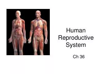
Human Reproductive System. Female Reproductive System. The main function of the female reproductive system is to produce ova; eggs cells.

E N D
Female Reproductive System • The main function of the female reproductive system is to produce ova; eggs cells. • The female reproductive structures will begin to develop and mature during puberty where the follicle-stimulating hormone (FSH) and lutenizing hormone (LH) are released from the hypothalamus and pituitary gland. These hormones activate the development of the sexual characteristics.
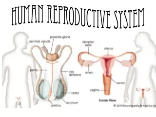
Eggs cells are produced in the ovaries. • The ovaries are located on either side of the uterus. • When an egg is mature each month, it is released into the fallopian tubes. These tubes are about 10 cm long and extend from the ovaries to the uterus.
The egg will travel through the fallopian tube until it reaches the uterus. • If the egg becomes fertilized it will attach to the wall of the uterus, where it will develop into an embryo. • An unfertilized egg will be broken down and discarded.
The uterus is about the size of a pear. It has three layers: an inner layer of epithelial, a middle layer of muscle and an outer layer of connective tissue. • The lower end of the uterus is called the cervix. • The cervix opens into the vagina, which is the birthing canal.
The main function of the male reproductive system is to produce sperm. • Males begin to produce sperm at puberty and continue throughout their lives.
Sperm production takes place in the testes. • Each testes produces millions of sperm. • Testosterone and FSH stimulate the production of sperm. • The testes are enclosed in a pouch called the scrotum.
The scrotum keeps the testes at a lower temperature then the rest of the body for production of sperm. • When immature sperm leave the testes, they travel to the epididymis, where they mature and remain until expelled.
During sexual stimulation, the sperm travel to the vas deferens. It is here where semen is produced. Fluids from the prostate gland, the bulbourethral gland secrete fluids that help protect the sperm and allow them to move more easily. • The seminal vesicle secretes fluids that help the sperm neutralize the acidity of the vagina.
During sexual arousal, semen flows from the vas deferens to the urethra. • When ejaculation occurs, there is a muscle that closes off the bladder to prevent urine from mixing with semen.
Egg Production & Menstruation • At birth females have about 2 million eggs that are partially developed. • Before puberty, most of the eggs will be broken down, and only about 400, 000 will be left. • At puberty, an increase in FSH, will stimulate an egg to complete meiosis I. One larger cell is developed and the other smaller cell, called the polar body, is discarded.
The larger cell will go through meiosis II after it becomes fertilized with a sperm cell. This cell develops into an egg, or ovum cell.
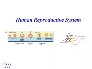
Each developing egg within the ovary is surrounded by a group of cells called a follicle. • When the egg is ready to be released, the follicle ruptures and the egg breaks through the ovary wall. • Ovulation, is the release of the egg.
After the egg leaves the ovary, it travels through the fallopian tube, where it can be fertilized by a sperm. • Within 5-7 days the egg moves from the fallopian tube to the uterus. • An unfertilized egg is disgarded during menstruation.
Menstrual Cycle • The menstrual cycle is a series of monthly changes within the reproductive system that includes producing and releasing an egg, and preparing the uterus to receive it. • The cycle is approximately 28 days and involves three stages.
Flow Phase • Day 1 of the cycle is the beginning of the menstrual flow. • During this phase the uterine lining detaches and is released, along with mucous, from the body.
Follicular Phase • This phase lasts from about day 6-14 • In the beginning estrogen is low. • The hypothalamus releases hormones which activate the pituitary gland to release FSH and LH. • This causes the egg and follicle to mature. • Ovulation occurs at about day 16.
When the egg is developing, it causes estrogen to increase. • An increase in estrogen leads to a thickening of the uterine lining. • At estrogen levels keep increasing, this leads to an increase in the production of LH. • The high levels of LH cause the follicle to rupture and release the egg.
Luteal Phase • After ovulation, the follicle turns yellow, and is now called the corpus luteum. • The corpus luteum releases estrogen and progesterone which causes the inhibition of LH. • The high levels of progesterone cause the uterine lining to thicken. There is also an increase in the number of blood vessels, in preparation for an embryo.
If an egg is not fertilized, then pituitary gland stops making FSH and LH. The corpus luteum breaks down, leading to the inhibition of progesterone and estrogen. • The inhibition of all the hormones leads to the shedding of the lining.
For most women the menstrual cycle lasts from pre-teen years until their mid 50s.
Production of Sperm • The production of sperm begins when the hypothalamus releases hormones that activate the pituitary gland to release FSH and LH. • FSH and LH will circulate to the testes which causes the testes to produce testosterone. • Production of testosterone will cause specialized cells to undergo meiosis and produce mature sperm.
The sperm cells will fully mature in the epididymis. • Each sperm has a head, tail, and midpiece. • The head contains a nucleus with 23 chromosomes as well as cap region called acrosome.
The acrosome releases enzymes that allow the sperm to penetrate the egg. • The midpiece contains mitochondria, which supplies the sperm with energy. • The tail acts to propel the sperm through the fallopian tube.
Process of fertilization • When sperm is released through ejaculation, they must swim through the uterus to the fallopian tubes. • When a sperm finds an eggs and binds, the sperm’s acrosome releases enzymes that digest the egg’s membrane. • Once one sperm enters, the membrane surface is changed so that no more sperm can enter.
After fertilization, the egg goes through meiosis II. • A zygote is formed when the 23 chromosomes from the sperm join with the 23 chromosomes of the egg.
Development of the Fetus • The zygote will go through mitosis as it starts to travel down the fallopian tube. • The zygote will continue to go through mitosis until it forms a hallow ball of cells known as a blastocyst. • The blastocyst will attach to the uterus wall.
Once attached, three layers of cells begin to develop: Ectoderm, mesoderm, and endoderm. • The ectoderm develops into the skin and nervous system. • The endoderm develops into the digestive system. • The mesoderm develops into the internal organs and tissues.
Once the structures begin to develop, the ball of cells is now called an embryo.
As the pregnancy continues, membranes will form around the embryo to protect it. The amnion membrane becomes fluid filled and is then called the amniotic sac.
The chorion membrane helps to nourish the embryo. • The chorion has small projections known as villi. These projections along with the uterus lining, form the placenta.
The placenta connects the mother to the fetus. It allows for exchange of nutrients, oxygen, and wastes. • The umbilical cord consists of two arteries and one vein. The cord connects the embryo to the placenta from the amniotic sac.
The placenta keeps the embryo’s and mother’s bloods separate. • Nutrients and oxygen from the mother diffuse into the chorionic villi and are then carried through the umbilical cord.
Zygote to Fetus Development • Human pregnancies are divided into three trimesters. • First trimester: • The heart begins beating at 5 weeks • Stem cells undergo differentiation into specialized cells and tissues • Embryo is susceptible to damage and mutations • Brain begins to develop • Weeks 1-8 the zygote is now called an embryo • At 9 weeks the embryo is called a fetus
Second Trimester: • Continued development and growth • At the end of the trimester, the fetus is about 30 cm long
Third Trimester: • Lungs develop completely • Fetus grows to about 50 cm