Download
1 / 46
711 likes | 1.55k Views
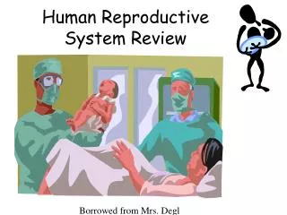
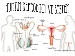
Human Reproductive System. Ch 36. SC.912.L.16.13 Describe the basic anatomy and physiology of the human reproductive system. Male. 1. Seminal vesicle 2. Prostate gland 3. Vas deferens 4. urethra 5. epididymis 6. scrotum 7. penis 8. testes. 1. Seminal vesicle.

E N D
SC.912.L.16.13 Describe the basic anatomy and physiology of the human reproductive system
Male • 1. Seminal vesicle • 2. Prostate gland • 3. Vas deferens 4. urethra • 5. epididymis • 6. scrotum • 7. penis • 8. testes.
1. Seminal vesicle • Elongated saccular organs with an irregular branching lumen. • Seminal vesicle fluid notable for high fructose and prostaglandin content.
2. Prostate gland • Weighs about 20 grams, with multiple excretory glands which empty into the urethra. Adds an alkaline fluid to the semen. • A normal human ejaculate has a volume of 2-5 cc and contains 150-200 million sperm.
3. Vas deferens • Sperm in the vas deferens is joined by seminal vesicle secretions as they pass through the prostate via the ejaculatory ducts into the urethra. • It is 35-45 cm long, and transports sperm. The vas provides rapid transport of sperm during ejaculation. It is what is “cut’ during a vasectomy.
4. urethra • The tube that carries both urine and semen out of the body. ( but not at the same time. )
5. epididymis The epididymis is a single convoluted duct approximately 20m long. It sits on top of the testes. The epididymis has several functions. 1 - Sperm tube: passage takes about 12 days in man, with sperm propelled by spontaneous rhythmic contractions of the duct. 2. Extra fluids are reabsorbed by the body. 3. Sperm storage 4. Sperm maturation.
6. scrotum • During the 8th fetal month the testis descend to the external position in the scrotum. The location is necessary for the lower temperature required for spermatogenesis
7. Penis • The penis is the male copulatory organ that has a main purpose of procreation by means of intercourse, but also serves as a part of the urinary system in males by excreting urine.
8. testes. • Made up of Seminiferous tubules the main function is to make sperm. • They also contain cells that produce testosterone, a male hormone. • Each tubule is 30-70 cm long and 200-300 um in diameter. There are approximately 500 tubules per testis. • Sperm produced by the seminiferous tubules pass out of the testis into the ductal system, and on into the epididymis. • In the adult, the testis acts as an exocrine organ, with the production and secretion of sperm. It also acts as an endocrine organ by its production and secretion of testosterone into the blood.
Sperm development Pseudo-colored scanning electron micrograph of seminiferous tubule.Purple cells are various stages of spermatocytes (cells that will give rise to sperm). They are surrounded by Sertoli cells in green, and the yellow cells at the center of the tube are spermatozoa that are ready to be on their way to the next organs, the epididymides (that’s plural for epididymis) Very COOL picture!!!!
Order of sperm leaving the body! SEVEN UP! Seminiferous tubule Epididymis Vas Deferens Ejaculatory ducts N (nothing) Urethra Penis
Female 1. Ovaries 2. oviduct ( fallopian tube) 3. uterus 4. cervix 5. vagina.
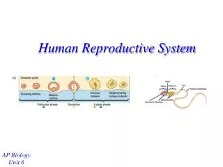
1. Ovaries • The ovaries are small, oval-shaped paired glands that are attached to each side of the uterus by a thin, fibrous ovarian ligament. The pair are responsible for storing and nurturing immature egg cells into mature eggs. Every month, one of them releases a mature egg into the Fallopian tube. • They also produce two main female sex hormones: the estrogen and progesterone, which are vital in regulating the menstrual cycles.
2. oviduct ( fallopian tube) • The Fallopian tubes are the two channels that connect the ovaries to the uterus. They are the location of fertilization. 1. Fimbriae: The fringe-like structure located at the end of the tube that captures egg released from the ovary and draws it into the tube. 2. Infundibulum: The funnel-like structure of the tube which is margined with fimbriae. 3. Ampulla: The longest portion of the tube with thin wall (almost muscle-free) and wide lumen. It is usually the portion where fertilization takes place. 4. Isthmus: The almost straight portion of the tube with relatively thick muscular wall and the narrowest lumen. 5. Interstitium: The portion of the tube that is closest to the uterus. It is sometimes known as the uterine portion of the tube for the fact that it lies within the uterus.
3. uterus • The uterus, or womb, is a hollow, pear-shaped organ with thick muscular wall; it lies in the middle of the pelvis, between the bladder and the rectum. The uterus is where implantation of the zygote occurs. It has a thick lining called the endometrium that contains nutrients for the fertilized egg, this lining is shed each month if a zygote is not implanted.
4. cervix • The cervix is the lower constricted segment of the uterus that joins with the upper part of vagina. The small cervical opening into the vagina is called external os while the one into the uterine cavity is called the internal os. They allow the sperm to enter the uterus during sexual intercourse and the menstrual fluid to flow out of the uterus during menstruation.
5. vagina. • The vagina is a muscular, narrow canal. It is also known as the birth canal. The inner wall of vagina is surfaced with numerous folds of soft elastic mucous membrane called vaginal rugae. They allow the vagina to expand considerably during sexual intercourse or childbirth. During menstruation, the vagina provides a channel for the menstrual fluid to flow out of the body.
Human Development • SC.912.L.16.13 Describe the process of human development from fertilization to birth and major changes that occur in each trimester of pregnancy. Ch 36
fertilization • The fertile period for a woman is 3 days before ovulation and 24 hours after. • Egg ( 23 chromosomes, haploid n) + sperm (23 chromosomes, haploid n) fuse to form the zygote ( 46 chromosomes diploid, 2n cell ). • This happens in the fallopian tube.
Immediately after fertilization the zygote goes through mitosis/cleavage (day 1) to form a mass of cells called a morula (day 4). It continues to divide until it forms a hollow ball of cells called a blastocyst ( days 5-9). The blastocyst then implants into the wall of the uterus, called the endometrium. ( about day 10)
placenta • Placenta: A temporary organ that joins the mother and fetus, transferring oxygen and nutrients from the mother to the fetus and permitting the release of carbon dioxide and waste products from the fetus. The placenta is roughly disk-shaped, and at full term it measures about 7 inches in diameter and slightly less than 2 inches thick.
Vanderbilt University Pictures • http://www.psy.vanderbilt.edu/courses/hon182/Stages_of_human_development.doc • The following slides are pictures and text from the above web site.
Week 2 • Fertilization, the joining of the sperm and the egg in the fallopian tube (below) to form a unique human being, occurs. Forty-six chromosomes provide the blueprint for the embryo’s physical characteristicThe picture on the right is a fertilized egg only thirty hours after conception. Magnified here, it is no larger than the head of a pin. Still rapidly dividing, the developing embryo, called a zygote at this stage, floats down from the fallopian tube and towards the uterus.
Week 5 • The embryo’s tiny heart begins to beat by day twenty-one. The brain has developed into 5 areas and some cranial nerves are visible. Arm and leg buds are visible and the formation of the eyes, lips, and nose has begun. The spinal cord grows faster than the rest of the body giving a tail like appearance which disappears as the embryo continues to grow. The placenta begins to provide nourishment for the embryo.
Week 7 • Major organs have all begun to form. The embryo has developed its own blood type, unique from the mother’s. Hair follicles and nipples form and knees and elbows are visible. Facial features are also observable. The eyes have a retina and lens. The major muscle system is developed and the embryo is able to move.
Week 8 • The embryo is reactive to its environment inside the amniotic sac where it swims and moves. Hands and feet can be seen. At the end of week 8, the embryonic period is over and the fetal stage begins.
Weeks 9-12 The heart is almost completely developed and the heart rate can be heard on a Doppler machine at the doctor’s office. Most major organs and tissues have developed and red blood cells are now produced in the liver. The face is well formed and the eyes are almost fully developed. The eyelids will close and not reopen until the 28th week. Arms, hands, fingers, legs, feet, and toes are fully formed. Nails and earlobes start to form and tooth buds develop in the gums. Fetus can make a fist with its finger. Testosterone (male sex hormone) is produced by the testes in male fetus.
Weeks 13-16 • The brain is fully developed and the fetus can suck, swallow, and make irregular breathing sounds. Fetus can feel pain (New England Journal of Medicine). Fetal skin is almost transparent. Muscles tissue is lengthening and bones are becoming harder. Liver and organs produce appropriate fluids. Eyebrows and eyelashes appear and the fetus makes active movements including kicks and even somersaults.
Week 20 • “Quickening” (when the mother can feel the fetus moving) usually occurs around this time. Finger and toenails appear. Lanugo, a fine hair now covers the entire body. The fetus can hear and recognize the mother’s voice. Sex organs are visible on ultrasound devices.
Week 24 • A protective waxy substance called Vernix covers the skin. By birth, most of the Vernix will be gone but any that is left is quickly absorbed. Fetus has a hand and startle reflex. Footprints and fingerprints are forming. Fetus practices breathing by inhaling amniotic fluid into its developing lungs.
Weeks 25 - 28 • Rapid brain development occurs during this period and the nervous system is able to control some bodily functions. The fetus’ eyelids now open and close. At 25 weeks there is a 60% chance of survival if born. The fetus is considered legally viable at 28 weeks and there is a 90% chance of survival if born at this point.
Weeks 29 - 32 • There is a rapid increase in the amount of body fat the fetus has. Rhythmic breathing occurs, but the lungs are not yet mature. The fetus sleeps 90-95% of the day. At this point there the survival rate is above 95% if the baby is born.
Weeks 38 - 40 • The fetus is considered full-term. Lanugo is gone except on upper arms and shoulders. Hair on the baby’s head is now coarser and thicker. The lungs are mature. The average weight of the baby at this point is seven and a half pounds. At birth the placenta detaches from the uterus and the umbilical cord will be cut as the baby takes his first breaths of air. Breathing will trigger changes in the heart and bypass arteries forcing all blood to now travel through the lungs.
http://www.psy.vanderbilt.edu/courses/hon182/Stages_of_human_development.dochttp://www.psy.vanderbilt.edu/courses/hon182/Stages_of_human_development.doc
Fertilization and the first trimester • Where does fertilization occur? In the fallopian tube (oviduct) • How many chromosomes (each) are in normal sperm and eggs? 23 3. What process occurs to make sure the eggs and sperm have the correct number of chromosomes? meiosis • What is a fertilized egg called? zygote 5. What process produces the ball of cells called a morula that implants into the uterus just a few days after fertilization? mitosis. 6. By the 10th day the hollow ball of cells called a blastocyst is fully implanted in the lining of the uterus called the what? Endometrium. 7. The placenta forms after 2 weeks. What is the main function of the placenta? To nourish and oxygenate the embryo 8. Organ systems begin to develop in the first trimester, the embryo changes its name at the end of the first trimester to now be called a what? Fetus
Second Trimester Basic changes: it can move and hear 1. Mostly a period of growth. The lungs are not yet fully developed. What starts to grow on the baby’s body? Hair, eyebrows and eyelashes 2. If the baby were to be born during the 2nd trimester, what might not be developed yet? Lungs and major organs
Third trimester Basic changes: rapid brain development, foot prints and finger prints, lungs continue to develop fat deposits form 1. Rapid weight gain and response to sound occurs. The bones are now fully developed. • What forms on the baby for insulation during the 3rd trimester? A layer of fat 3. What are now fully developed and ready for birth after the 3rd trimester? Lungs