Download
1 / 35
350 likes | 1.03k Views
D- Fatty Acid Synthesis. The input to fatty acid synthesis is acetyl-CoA , which is carboxylated to malonyl-CoA. ATP-dependent carboxylation provides energy input. The CO 2 is lost later during condensation with the growing fatty acid.

E N D
The input to fatty acid synthesis is acetyl-CoA, which is carboxylated to malonyl-CoA. ATP-dependent carboxylation provides energy input. TheCO2 is lost later during condensation with the growing fatty acid. The spontaneous decarboxylation drives the condensation reaction.
Acetyl-CoA Carboxylase catalyzes the 2-step reaction by which acetyl-CoA is carboxylated to form malonyl-CoA. As with other carboxylation reactions, the enzyme prosthetic group is biotin. ATP-dependent carboxylation of the biotin, carried out at one active site 1 , is followed by transfer of the carboxyl group to acetyl-CoA at a second active site 2 .
The overall reaction, which is spontaneous, may be summarized as: HCO3− +ATP+ acetyl-CoAàADP+ Pi + malonyl-CoA
Biotin is linked to the enzyme by an amide bond between the terminal carboxyl of the biotin side chain and the ε-amino group of a lysine residue. The combined biotin and lysine side chains act as a long flexible arm that allows the biotin ring to translocate between the 2 active sites.
Acetyl-CoA Carboxylase, which converts acetyl-CoA to malonyl-CoA, is the committed step of the fatty acid synthesis pathway. The mammalian enzyme is regulated, by • phosphorylation • allosteric control by local metabolites.
Conformational changes associated with regulation: • In the active conformation, Acetyl-CoA Carboxylase associates to form multimeric filamentous complexes. • Transition to the inactive conformation is associated with dissociation to yield the monomeric form of the enzyme (protomer).
AMP functions as an energy sensor and regulator of metabolism. When ATP production does not keep up with needs, a higher portion of a cell's adenine nucleotide pool is in the form of AMP. • AMPpromotes catabolic pathways that lead to synthesis of ATP. • AMPinhibits energy-utilizing synthetic pathways. E.g., AMP regulates fatty acid synthesis and catabolism by controlling availability of malonyl-CoA.
AMP-Activated Kinase catalyzes phosphorylation of Acetyl-CoA Carboxylase. This causes inhibition of ATP-utilizing of malonyl-CoA production. • Fatty acid synthesis is diminished by lack of the substrate malonyl-CoA. • As discussed earlier, fatty acid oxidation is stimulated due to decreased inhibition by malonyl-CoA of transfer of fatty acids into mitochondria.
result in phosphorylation of Acetyl-CoA Carboxylase via cAMP-Dependent Protein Kinase. With Acetyl-CoA Carboxylase inhibited, acetyl-CoA remains available for synthesis of ketone bodies, the alternative metabolic fuel used when blood glucose is low. A cAMP cascade, activated by glucagon & epinephrine when blood glucose is low, may also
The antagonistic effect of insulin, produced when blood glucose is high, is attributed to activation of Protein Phosphatase.
Palmitoyl-CoA (product of Fatty Acid Synthase) promotes the inactive conformation, diminishing production of malonyl-CoA, the precursor of fatty acid synthesis. This is an example of feedback inhibition. Regulation of Acetyl-CoA Carboxylase by local metabolites:
[Citrate] is high when there is adequate acetyl-CoA entering Krebs Cycle. Excess acetyl-CoA is then converted via malonyl-CoA to fatty acids for storage. Citrate allosterically activates Acetyl-CoA Carboxylase.
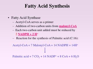
Fatty acid synthesis from acetyl-CoA & malonyl-CoA occurs by a series of reactions that are: • in bacteria catalyzed by 6 different enzymes plus a separate acyl carrier protein (ACP) • in mammals catalyzed by individual domains of a very large polypeptide that includes an ACP domain. Evolution of the mammalian Fatty Acid Synthase apparently has involved gene fusion. NADPH serves as electron donor in the two reactions involving substrate reduction. The NADPH is produced mainly by the Pentose Phosphate Pathway.
Fatty Acid Synthase prosthetic groups: • the thiol of the side-chain of a cysteine residue of Condensing Enzyme domain. • the thiol of phosphopantetheine, equivalent in structure to part of coenzyme A.
Phosphopantetheine (Pant) is covalently linked via a phosphate ester to a serine OH of the acyl carrier protein domain of Fatty Acid Synthase. Thelong flexible arm of phosphopantetheine helps its thiol to move from one active site to another within the complex.
As each of the substrates acetyl-CoA & malonyl-CoA bind to the complex, the initial attacking group is the oxygen of a serinehydroxyl group of the Malonyl/acetyl-CoA Transacylase enzyme domain. Each acetyl or malonyl moiety is transiently in ester linkage to this serine hydroxyl, before being transferred into thioester linkage with the phosphopantetheine thiol of the acyl carrier protein (ACP) domain. Acetate is subsequently transferred to a cysteine thiol of the Condensing Enzyme domain.
The condensation reaction (step 3) involves decarboxylation of the malonyl moiety, followed by attack of the resultant carbanion on the carbonyl carbon of the acetyl (or acyl) moiety.
The β-ketone is reduced to an alcohol by e− transfer from NADPH. • Dehydration yields a trans double bond. • Reduction by NADPH yields a saturated chain.
Following transfer of the growing fatty acid from phosphopantetheine to the Condensing Enzyme's cysteine sulfhydryl, the cycle begins again, with another malonyl-CoA.
Product release: When the fatty acid is 16 carbon atoms long, a Thioesterase domain catalyzes hydrolysis of the thioester linking the fatty acid to phosphopantetheine. The 16-C saturated fatty acidpalmitateis the final product of the Fatty Acid Synthase complex.
The primary structure of the mammalian Fatty Acid Synthase protein is summarized above. Fatty Acid Synthase in mammals is a homo-dimer. X-Ray crystallographic analysis at 3.2 Å resolution shows the dimeric Fatty Acid Synthase to have an X-shape. The 2 copies of the protein are displayed at right in different colors.
The domain arrangement is shown below. Each copy of the dimeric protein has an S shape, with the N-terminalKS (Condensing Enzyme / β-Ketoacyl Synthase) domain folded back to form part of the central interaction domain. KR = β-Ketoacyl Reductase; ER = Enoyl Reductase; DH = Dehydratase; KS = β-Ketoacyl Synthase (Condensing Enzyme); MAT = Malonyl/Acetyl-CoA Transacylase.
The X-ray analysis does not resolve the C-terminal ACP (acyl carrier protein) & Thioesterase domains, predicted from the primary structure to be near the KR domains. These domains may be too flexible to be resolved by crystallography. KR = β-Ketoacyl Reductase; ER = Enoyl Reductase; DH = Dehydratase; KS = β-Ketoacyl Synthase (Condensing Enzyme); MAT = Malonyl/Acetyl-CoA Transacylase.
Fatty Acid Synthase complex is somewhat asymmetric. There is evidence for conformational changes relating to catalysis. Protein flexibility may facilitate transfer of ACP-attached reaction intermediates among the several active sites in each half of the complex.
Explore with Chime the structure of the Mammalian Fatty Acid Synthase III.
1 1 Palmitate, a 16-C saturated fatty acid,is the final product of the Fatty Acid Synthase reactions. 1. a. How many acetyl-CoA used for initial priming of enzyme? _____ b. How many acetyl-CoA used for synthesis of each malonate? ____ c. How many malonate used (how many reaction cycles) per synthesis of one 16-C palminate? ________ d. Total acetyl-CoA used for priming & for syntheisis of malonate, a + b(c): ________ 2. a. How many ~P bonds of ATP used for synthesis of each malonate? ________ b. Total ~P bonds of ATP used for synthesis of one 16-C palmitate, 2a(1c): ________ 3. a. How many NADPH used per reaction cycle? __________ b. Total NADPH used per synthesis of one 16-C palmitate, 3a(1c): _________ 7 8 1 7 2 14
Summary (ignoring H+ & water): Write a balanced equation for synthesis of palmitate from acetyl-CoA, listing net inputs and outputs: 8acetyl-CoA + 14NADPH + 7ATPà palmitate + 14NADP++ 8 CoA+7ADP+7Pi Summary based on malonate as an input: acetyl-CoA + 7malonyl-CoA + 14NADPHà palmitate + 7CO2 + 14NADP+ + 8CoA Fatty acid synthesis occurs in the cytosol. Acetyl-CoA generated in mitochondria is transported to the cytosol via a shuttle mechanism involving citrate.
Fatty Acid Synthase is transcriptionally regulated. In liver: • Insulin, a hormone produced when blood glucose is high, stimulates Fatty Acid Synthase expression. Thus excess glucose is stored as fat. Transcription factors that that mediate the stimulatory effect of insulin include USFs (upstream stimulatory factors) and SREBP-1. SREBPs (sterol response element binding proteins) were first identified for their regulation of cholesterol synthesis. • Polyunsaturated fatty acidsdiminish transcription of the Fatty Acid Synthase gene in liver cells, by suppressing production of SREBPs.
In fat cells: Expression of SREBP-1 and of Fatty Acid Synthase is inhibited by leptin, a hormone that has a role in regulating food intake and fat metabolism. Leptin is produced by fat cells in response to excess fat storage. Leptin regulates body weight by decreasing food intake, increasing energy expenditure, and inhibiting fatty acid synthesis.
Elongation beyond the 16-C length of the palmitate product of Fatty Acid Synthase is mainly catalyzed by enzymes associated with the endoplasmic reticulum (ER). ER enzymes lengthen fatty acids produced by Fatty Acyl Synthase as well as dietary polyunsaturated fatty acids. Fatty acids esterified to coenzyme A serve as substrates. Malonyl-CoA is the donor of 2-carbon units in a reaction sequence similar to that of Fatty Acid Synthase except that individual steps are catalyzed by separate proteins. A family of enzymes designated Fatty Acid Elongases or ELOVL (elongation of very long chain fatty acid) catalyze the initial condensation step.
Desaturases introduce double bonds at specific positions in a fatty acid chain. Mammalian cells are unable to produce double bonds at certain locations, e.g., Δ12. Thus some polyunsaturated fatty acids are dietary essentials, e.g., linoleic acid, 18:2 cis Δ9,12 (18 C atoms long, with cis double bonds at carbons 9-10 & 12-13).
Formation of a double bond in a fatty acid involves the following endoplasmic reticulum membrane proteins in mammalian cells: • NADH-cyt b5 Reductase, a flavoprotein with FAD as prosthetic group. • Cytochrome b5, which may be a separate protein or a domain at one end of the desaturase. • Desaturase, with an active site that contains two iron atoms complexed by histidine residues.
The desaturase catalyzes a mixed function oxidation reaction. There is a 4-electron reduction of O2à2 H2O as a fatty acid is oxidized to form a double bond. • 2e− pass from NADH to the desaturase via the FAD-containing reductase & cytochrome b5, the order of electron transfer being: NADHàFADàcyt b5àdesaturase • 2e−are extracted from the fatty acid as the double bond is formed. E.g., the overall reaction for desaturation of stearate (18:0) to form oleate (18:1 cis Δ9) is: stearate + NADH + H+ + O2àoleate + NAD+ + 2H2O