Download
1 / 79
790 likes | 992 Views
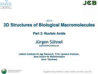
- 2010 - 3D Structures of Biological Macromolecules Part 3: Nucleic Acids. Jürgen Sühnel jsuehnel@fli-leibniz.de. Leibniz Institute for Age Research, Fritz Lipmann Institute, Jena Centre for Bioinformatics Jena / Germany. Supplementary Material: www.fli-leibniz.de/www_bioc/3D/.

E N D
- 2010 - 3D Structuresof Biological Macromolecules Part 3: Nucleic Acids Jürgen Sühnel jsuehnel@fli-leibniz.de Leibniz Institute for Age Research, Fritz Lipmann Institute, Jena CentreforBioinformatics Jena / Germany Supplementary Material: www.fli-leibniz.de/www_bioc/3D/
Nucleic Acids DNA genomic information (nucleosomes, chromatin) RNA messenger RNA, ribosomal RNA, transfer RNA, ribozymes, small RNAs, noncoding RNAs, RNAi (gene silencing), aptamers (alternatives to antibodies) PNA peptide nucleic acids mimic nucleic acids
New Roles for RNA Couzin J. Breakthrough of the year. Small RNAs make big splash.Science.2002, 298, 2296-2297.
Chain Direction in Nucleic Acids /web.siumed.edu/~bbartholomew/course_material/nucleic_acids.html; no longer active)
Nucleic Acid Base Pairs The ten possible purine-pyrimidine base pairs. Source: Ignacio Tinoco, Jr. in Gesteland, R. F. and Atkins, J. F. (1993) THE RNA WORLD. Cold Spring Harbor Laboratory Press.
Nucleic Acid Base Pairs The seven possible homo-purine base pairs. Source: Ignacio Tinoco, Jr. in Gesteland, R. F. and Atkins, J. F. (1993) THE RNA WORLD. Cold Spring Harbor Laboratory Press.
Nucleic Acid Base Pairs The four possible hetereo-purine base pairs. Source: Ignacio Tinoco, Jr. in Gesteland, R. F. and Atkins, J. F. (1993) THE RNA WORLD. Cold Spring Harbor Laboratory Press.
Nucleic Acid Base Pairs The seven possible pyrimidine-pyrimidine base pairs.
DNA Hydration www.lsbu.ac.uk/water/nucleic.html
Non-Canonical Base Pair Database http://prion.bchs.uh.edu/bp_type/
Base Triples in tRNA Base triads in the crystal structure of yeast phenylalanine transfer RNA (PDB code: 4tna).
Base Tetrads in a DNA Tetraplex Parallel-stranded DNA tetraplex formed from the Tetrahymena telomeric sequence d(TTGGGGT) solved by NMR spectroscopy (PDB code: 139d). The structure contains four stacked G-tetrads in the center and additional T-tetrads.
Base Polyads Base triads and a heptad in the crystal structure of a pseudoknot from beet western yellow virus (BWYV) involved in frameshifting (PDB code: 437d). The heptad is formed from two triads linked by A25.
HBexplore – H-bond Analysis in Proteins and Nucleic Acids www.imb-jena.de/www_bioc/hbx/hbx.html
Geometrical Parameters for Base Pairs in Nucleic Acids ndbserver.rutgers.edu/archives/report/tsukuba_sup/bp_step_hel.html
Nucleic Acid Structure P: P is the pitch of the helix corresponding to the distance between a base and the base obtained after walking along the DNA one full turn of 360°. n: n is the number on nucleotides within one pitch. h: distance between base planes. online-media.uni-marburg.de/chemie/bioorganic/vorlesung1/kapitel1e.html?/chemie/bioorganic/vorlesung1/k1e-20.html
DNA Conformations A B Z www.rcsb.org/pdb/molecules/pdb23_3.html
Nucleic Acid Conformations • B-DNA is found at low salt concentrations. It is believed to be the native conformation occurring in chromatin. • In the cell nucleus DNA is complexed with about an equivalent mass of protein to form a structure • known as chromatin. Chromatin is a periodic structure made up of repeating, regularly spaced • subunits, the subunit being the nucleosome. Within the nucleosomes the major part of DNA is • wrapped around histones. The remaining DNA joining each nucleosome is known as linker DNA. • A-DNA In solutions with higher salt concentrations or with alcohol added A-DNA is found. • Z-DNA occurs for alternating poly(dG-dC) sequences in solutions with high salt concentrations or alcohol. • RNA occurs (contrary to DNA) almost exclusively in the A-conformation (or in a related A'-form). • There are further nucleic acid conformations like C-DNA, H-DNA or others which are not discussed • here. • Geometrical features: • The distance between two subsequent base pairs along the helical axis is called helical rise (h). • The pitch (p) is the length of the helix axis for one complete helix turn. • The turn angle per nucleotide or twist angle (t) is given by 360° / number of nucleotides per turn. • C2'-endo and C3'-endo are descriptions of sugar conformations. • The most frequently occurring nucleic acid model conformations are characterized by the following geometrical • parameters : • A-DNA • right-handed helix; sugar pucker: C3'-endo; number of nucleotides per pitch: 11; h: 2.56 Å; t: +32.7°. • B-DNA • right-handed helix; sugar pucker: C2'-endo; number of nucleotides per pitch: 10; h: 3.38 Å; t: +36° . • Z-DNA • left-handed helix; G: syn conformation; sugar pucker: C3'-endo; C: anti conformation, sugar pucker: C2' • endo; number of nucleotides per pitch: 6x2; h: 3.7x2 Å; t= -30°x2 (for Z-DNA the repeat unit is the dimer • (G-C)2.
First Single-crystal DNA Structure (B-DNA) Drew-Dickerson structure H. R. Drew, R. M. Wing, T. Takano, C. Broka, S. Tanaka, K. Itakura, R. E. Dickerson Structure Of A B-/DNA Dodecamer. Conformation And Dynamics Proc. Nat. Acad. Sci. Usa V. 78 2179 1981