Download
1 / 66
660 likes | 902 Views
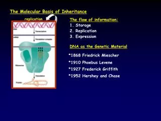
Chapter 16. The molecular basis of heredity. DNA: The Genetic Material. By the early twentieth century, geneticists had associated the presence of genes with chromosomes. Circumstantial evidence pointed to DNA as the genetic material.

E N D
Chapter 16 The molecular basis of heredity
DNA: The Genetic Material • By the early twentieth century, geneticists had associated the presence of genes with chromosomes. • Circumstantial evidence pointed to DNA as the genetic material. • DNA was found in the nucleus and chromosomes, which were already known to carry genes. • A dye that binds to DNA showed that the amount of DNA in somatic cells was twice that in eggs or sperm, as would be expected from Mendel’s discoveries.
DNA: The Genetic Material • In the 1920s, the English physician Frederick Griffith did experiments with two strains of Streptococcus pneumoniae. • He discovered that a chemical transforming principle from one strain could could cause a heritable change in the other strain.
Figure 16.1 Genetic Transformation of Nonvirulent Pneumococci
DNA: The Genetic Material • Oswald T. Avery and colleagues spent several years identifying the transforming principle. • They did cell fractionation in order to separate out the different components. • They treated the extract from the S-strain bacteria in various ways to destroy different types of substances but retain others. • When DNA was destroyed, the transforming activity was lost, but when DNA was left intact, the transforming activity survived.
DNA: The Genetic Material • In 1952, Alfred D. Hershey and Martha Chase performed experiments confirming that DNA is the genetic material. • The T2 bacteriophage, a virus that attacks E. coli, consists almost entirely of a DNA core packed in a protein coat. • When a T2 bacteriophage attacks a bacterium, part but not all of the virus enters the bacterial cell. • The Hershey-Chase experiment determined which part of the virus (protein or DNA) entered the bacterium.
Figure 16.2 T2 and the Bacteriophage Reproduction Cycle (Part 1)
Figure 16.2 T2 and the Bacteriophage Reproduction Cycle (Part 2)
DNA: The Genetic Material • Some viruses were labeled with radioactive sulfur, which is present in proteins but not in DNA. • Other viruses were labeled with radioactive phosphorus, which is present in DNA but absent from most proteins. • The labeled sulfur (and thus the viral protein) separated from the bacteria, but the labeled phosphorus (and thus the viral DNA) remained with the bacteria.
The Structure of DNA • Scientists set out to determine the structure of DNA hoping to find the answers to two questions: • How is DNA replicated between nuclear divisions? • How does DNA cause the synthesis of specific proteins? • The structure of DNA was determined after many types of evidence were combined.
The Structure of DNA • The positions of atoms in a crystalline substance can be inferred from the pattern of diffraction of X-rays passed through it. • In the early 1950s, many skilled X-ray crystallographers tried but failed to glean information from X-ray diffraction patterns of DNA. • The English chemists Rosalind Franklin and Maurice Wilkins were able to provide key information about the structure of DNA based on X-ray crystallography.
Figure 16.4 X-Ray Crystallography Revealed the Basic Helical Structure of the DNA Molecule
The Structure of DNA • By the 1950s it was known that DNA was a polymer of nucleotides. • The four nucleotides that make up DNA differ only in their nitrogenous bases. • There are two purines (adenine and guanine) and two pyrimidines (cytosine and thymine). • In 1950, Erwin Chargaff noted that in DNA from all species tested, the amount of adenine equals the amount of thymine, and the amount of guanine equals the amount of cytosine.
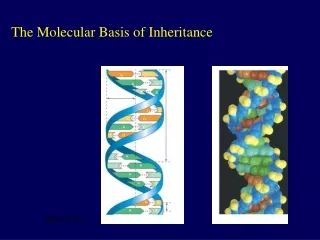
The Structure of DNA • English physicist Francis Crick and American geneticist James D. Watson established the general structure of DNA. • The results of X-ray crystallography convinced them that the DNA molecule was helical. • X-ray crystallography also provided the values of certain distances within the helix. • Density measurements and earlier models pointed to a structure with two polynucleotide chains running antiparallel to each other.
The Structure of DNA • Four features summarize the molecular architecture of DNA: • The DNA molecule is a double-stranded helix. • The diameter of the DNA molecule is uniform. • The twist in DNA is right-handed. • The two strands run in different directions (they are antiparallel).
The Structure of DNA • The sugar–phosphate backbones of each strand coil around the outside of the helix. • The nitrogenous bases point toward the center of the helix. • Hydrogen bonds between complementary bases hold the two strands together. • A always pairs with T (two hydrogen bonds). • G always pairs with C (three hydrogen bonds).
The Structure of DNA • The phosphate groups link the 3¢carbon of one deoxyribose molecule to the 5¢ carbon of the next. • Thus a single strand of DNA has a 5¢ phosphate group at one end (the 5¢ end) and a free 3¢hydroxyl group at the other end (the 3¢end). • In a double helix, the 5¢ end of one polypeptide is hydrogen-bonded to the 3¢end of the other, and vice versa.
The Structure of DNA • The genetic material performs four important functions: • It stores all of an organism’s genetic information. • It is susceptible to mutation. • It must be precisely replicated in the cell division cycle. • It is expressed as the phenotype.
Determining the DNA Replication Mechanism • American biochemist Arthur Kornberg demonstrated that the DNA molecule contains the information needed for its own replication. • Kornberg showed that DNA can replicate in a test tube with only a specific enzyme (DNA polymerase) and a mixture of four precursors (deoxyribonucleoside triphosphates): dATP, dCTP, dGTP, and dTTP.
Determining the DNA Replication Mechanism • Theoretically, DNA could serve as its own template in one of three different ways: • Semiconservative replication would use each parent strand as a template for a new strand. • Conservative replication would build an entirely new double helix based on the template of the old double helix. • Dispersive replication would use fragments of the original DNA molecule as templates for assembling two molecules.
Determining the DNA Replication Mechanism • Matthew Meselson and Franklin Stahl demonstrated in 1957 that DNA replication is semiconservative by using a technique called density labeling. • They used DNA labeled with “heavy” nitrogen (15N).
The Molecular Mechanisms of DNA Replication • DNA replication takes place in two steps: • The hydrogen bonds between the two strands are broken, making each strand available for base pairing. • The new nucleotides are covalently bonded to each growing strand.
The Molecular Mechanisms of DNA Replication • In DNA replication, nucleotides are added to the 3¢ end of the growing strand. • The three phosphate groups of the deoxyribonucleoside triphosphate are attached to the 5¢ position of the sugar. • Energy for synthesis of nucleotides to the growing chain comes from breaking the bonds between these three phosphates.
Figure 16.10 Each New DNA Strand Grows from its 5¢ End to its 3¢ End
The Molecular Mechanisms of DNA Replication • A huge protein complex catalyzes DNA replication. • This replication complex recognizes an origin of replication on a chromosome. • DNA replicates in both directions from the origin, forming two replication forks. • In DNA replication, both strands of DNA act as templates. • Recent evidence suggests that the replication complex is stationary, and DNA threads through it.
The Molecular Mechanisms of DNA Replication • The enzyme DNA helicase uses energy from ATP to unwind the two DNA strands. • Special proteins bind to the unwound strands to keep them apart. • Small chromosomes, such as those found in bacteria, have a single origin of replication. • Replication in bacteria produces two interlocking circular DNAs that are separated by the enzyme DNA topoisomerase.
Figure 16.12 (a)Replication in Small Circular and Large Linear Chromosomes
The Molecular Mechanisms of DNA Replication • Large chromosomes can have hundreds of origins of replication. • Replication occurs at many different sites simultaneously.
Figure 11.12 (b)Replication in Small Circular and Large Linear Chromosomes
The Molecular Mechanisms of DNA Replication • DNA polymerases cannot build a strand without having an existing strand, called a primer, to start from. • In DNA replication, the primer strand is a short strand of RNA complementary to the DNA template strand. • An enzyme called a primase makes the primer strand. • The primase is part of a protein complex called a primosome.
The Molecular Mechanisms of DNA Replication • Most cells contain more than one DNA polymerase. • Only one of the polymerases is responsible for chromosomal DNA replication. • The others are involved in primer removal and DNA repair.
Figure 11.15 Many Proteins Collaborate at the Replication Fork
The Molecular Mechanisms of DNA Replication • Recall that new bases are always added to the 3¢end of a growing DNA strand. • The strands in the template DNA are antiparallel, however. • As a result, as the strands pass through the replication complex, one strand (the leading strand) will be in the correct orientation for addition of new nucleotides. • The other strand (the lagging strand) will be in the reverse orientation.
The Molecular Mechanisms of DNA Replication • Because of its backward orientation, the lagging strand must grow in relatively small, discontinuous pieces, called Okazaki fragments. • Each Okazaki fragment requires an RNA primer strand, which is formed by RNA primase. • DNA polymerase III synthesizes complementary DNA starting from the 3¢end of the new primer and working toward the previous Okazaki fragment.
The Molecular Mechanisms of DNA Replication • When DNA polymerase III reaches the previous Okazaki fragment, it is released. • DNA polymerase I then replaces the RNA primer of the previous Okazaki fragment with DNA. • Finally, DNA ligase catalyzes formation of the phosphodiester linkage that joins the two Okazaki fragments.
The Molecular Mechanisms of DNA Replication • Recall that replication of the lagging strand occurs by the addition of Okazaki fragments to RNA primers. • Beyond the very end of a linear DNA molecule, there is no place for a primer to bind. • New chromosomes formed after DNA replication have single-stranded DNA at each end. • This single-stranded region is cut off, slightly shortening the chromosome after each cell division.
The Molecular Mechanisms of DNA Replication • Many eukaryotic chromosomes have repetitive sequences called telomeres at their ends that shorten after each round of cell division. • After a given number of cell divisions, the telomeres have shortened to the extent that they are no longer able to stabilize the ends of the chromosomes, and no cell division can occur. • This results in cell death and explains in part why cells do not last the entire lifetime of the organism.
The Molecular Mechanisms of DNA Replication • Constantly dividing cells, such as bone marrow, germ line, and more than 90 percent of cancer cells, produce an enzyme called telomerase that catalyzes the addition of any lost telomeric sequences.