Download
1 / 1
10 likes | 155 Views
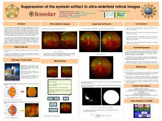
State of the Art. (a) (b). Figure 2. Original images. 45 deg. 200 deg. Suppression of the eyelash artifact in ultra-widefield retinal images. Dr. Delia Cabrera – dcabrera2@med.miami.edu.

E N D
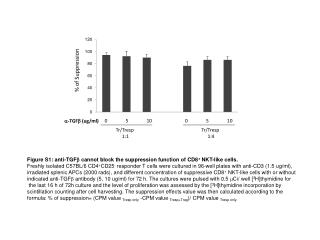
State of the Art (a) (b) Figure 2. Original images 45 deg 200 deg Suppression of the eyelash artifact in ultra-widefield retinal images Dr. Delia Cabrera – dcabrera2@med.miami.edu Vanessa Ortiz-Rivera – ortiz@rpi.edu Dr. Badrinath Roysam, Advisor – roysam@ece.rpi.edu Dr. Charles Stewart – stewart@cs.rpi.edu Gehua Yang – yangg2@rpi.edu Conclusions Abstract Ultra-widefield images Experimental Results Retinal imaging is used by optometrists and ophthalmologists to screen for and diagnose eye and non-eye diseases. It is believed that indicators of systemic diseases and precursors to different eye conditions often exhibit first on the periphery of the retina. Current retinal examination methods provide a narrow field of view (about 5%) and can therefore miss eye and non-eye disorders that may be present and can be detected on the periphery of the retina. However as the field of view becomes greater, certain artifacts can be captured in the image making its analysis more challenging. An example of this is the presence of eyelashes. In this work, an automated image-pair registration method known as the Generalized Dual-Bootstrap Iterative Closest Point (GDB-ICP) algorithm [2]-[4], was used to suppress the eyelash artifact, of ultra-widefield retinal images. The percent of suppression evidenced in the mosaic created by the algorithm was quantified. For the pair of images used in this work the percent of suppression obtained, was 6.13% in regards to the overall image. • GDB-ICP image registration algorithm successfully registered the set of images used for this work. • From preliminary results, the algorithm seems to be robust to the presence of eyelashes in the images. • The mosaic created from the transformed images shows a degree of suppression of the eyelash artifact, which was quantified to be 6.13% in regards to the overall image. • Further analysis should be performed by using new data sets in order to validate the results presented. A) The following images were obtained as a result of the image registration stage: The following pair of images were analyzed in this work: Acknowledgments • Current analysis of eye conditions is been done with images taken over a field of view of 30º. • To capture a greater percentage of the retina, either the patient's eye must be dilated, causing patient discomfort, or multiple images of the retina must be taken, at additional cost and time to the practitioner [1]. This work was supported in part by Gordon-CenSSIS, the Bernard M. Gordon Center for Subsurface Sensing and Imaging Systems, under the Engineering Research Centers Program of the National Science Foundation (Award Number EEC-9986821). The GDB-ICP image pair registration algorithm was developed by the Computer Vision research group led by Dr. Charles Stewart, professor of the Department of Computer Science at RPI. Special thanks to Dr. Delia Cabrera, from Bascom Palmer Eye Institute at the University of Miami, for providing us with the data used in this work. (a) (b) • The red circles are used to illustrate the different positions of a common feature among the images. The misalignment (represented by the line’s angle) as well as the presence of the eyelash artifact are evident. Figure 3. Registered images Optomap ® Instrument Methodology • Optomap® is the core product of the company Optos ® and generates a digital wide-field (200 degrees internal angle) image of the retina. • Image capture takes a quarter of a second once the patient is positioned relative to the device. • The device is designed to be able to take an image through a 2mm aperture, and therefore the dilation is not necessary. References [1] http://www.optos.com/ [2] Gehua Yang, Charles V. Stewart, Michal Sofka, and Chia-Ling Tsai, "The Generalized Dual-Bootstrap ICP algorithm with application to registering challenging image pairs”, IEEE Transactions on Pattern Analysis and Machine Intelligence [3] Gehua Yang, Charles V. Stewart, Michal Sofka, Chia-Ling Tsai: “Automatic robust image registration system: Initialization, estimation, and decision”. Proceedings of the IEEE International conference on Computer Vision Systems (ICVS), pp. 23-31, 2006. [4] C.V. Stewart, C.-L. Tsai and B. Roysam, “The Dual-Bootstrap Iterative Closest Point algorithm with application to retinal image registration”, IEEE Trans. on Medical Imaging , October 2003. Image1 Image2 Figure 4. Mosaic obtained from registered images. GDB-ICP algorithm: Step 1: Initialization- extraction of keypoints from Image1 and Image2. Step 2: Estimation of transformation parameters. Step 3: Decision making – determines if an estimate generated by the algorithm is a correct alignment of the two images. Figure 1. Picture of the optomap® imager B) The percent of eyelash suppression exhibited in the mosaic, was quantified by comparing both Image1t and Image2t. As a result a mask was obtained. In Figure 5(a), white pixels represent areas from which the eyelashes were suppressed. Conventional Retinal Imaging: Technology only captures a small area of the retina at one time. Contact Information Dr. Badrinath Roysam, Professor Department of Electrical, Computer and Systems Engineering Associate Director, NSF Center for Subsurface Sensing & Imaging Systems (CenSSIS ERC) Rensselaer Polytechnic Institute 110 8th Street, Troy, New York 12180-3590. Office(JEC 7010): 518-276-8067, Lab(JEC 6308): 518-276-8207, Fax: 518-276-8715 Email: roysam@ecse.rpi.edu, Web: http://www.ecse.rpi.edu/~roysam Output Size: 1200 x 820 Image1t Image2t Mosaic Value Added to CenSSIS Figure 2. Field of view of conventional retinal imaging. Retinal Imaging with Optomap ® : The majority of the retina is captured with a single image. Masking (a) (b) Figure 5. (a) Area of eyelash suppression. (b) Overall image area. Compare and calculate suppression The percent of suppression was calculated from the ratio of white pixels on mask (a) and white pixels on mask (b). This is: Image1t, Image2t: Transformed images Figure 3. Field of view with optomap® . Size: 3900 x 3072