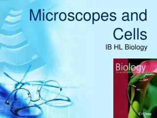
Microscopes and Cells
Microscopes and Cells. IB HL Biology. 2.1.4 Comparison of relative sizes of molecules, cell membrane thickness, viruses, bacteria, organelles and cells, using the appropriate SI unit. Refer Figure 1.18, Clegg p20 Size relationships of biological and chemical

Microscopes and Cells
E N D
Presentation Transcript
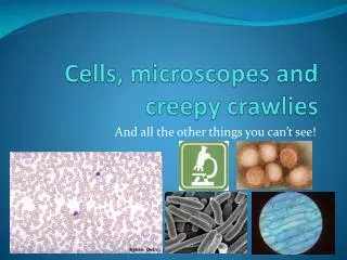
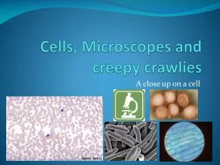
Microscopes and Cells IB HL Biology
2.1.4 Comparison of relative sizes of molecules, cell membrane thickness, viruses, bacteria, organelles and cells, using the appropriate SI unit. Refer Figure 1.18, Clegg p20 Size relationships of biological and chemical levels of organization are compared -Notice the diversity and how the power of 10 is used -Although sizes are expressed in length and diameter, cells and organisms are 3-D
Molecules: 1 nm Membranes (on organelles): 10 nm Viruses: 100 nm Bacteria: 1 um Organelles: up to 10 um Most cells: up to 100 um Measurements above in 2D, remember all structures have 3D shape. Size of various cells and structures:
Kilo- 1000 Units Hecto- 100 units Dek- 10 units Multiply Basic Unit Deci- 0.1 units Divide Centi- 0.01 units Milli- 0.001 units The Metric System • Know how to convert from one unit to another.
What units are used to measure cells? • 1 mm = 1000 micrometers (um) • 1 mm = 1,000,000 nanometers (nm) • Or… • A micrometer is 1 x 10⁻³ mm (0. 001) • A nanometer is 1 x 10⁻⁶ mm (0.000001 mm)
Magnification and Scale bars • Specimen size how large the specimen actually is • Image size how large the specimen appears in a drawing or photograph • Magnification how much larger the image is than the actual size • Formula used for these calculations: Magnification = size of image size of specimen Microscopy CalculationsYoutube video
Calculating Linear Magnification • What is the actual size of this specimen in um? • 60mm/5 = 12mm • 12mm x 1000 um = • 12,000 um Magnification x5 60 mm Measuring picture
2.1.5 Calculate the linear magnification of drawings and the actual size of specimens in images of known magnification Magnification could be stated (for example, ×250) or indicated by means of a scale bar Magnification = Image size = 58mm = 2.5mm x 1000 = x23200 Size of specimen 23mm(size of specimen meas with scale bar) Scale bar 1um Refer to image on p16 and problem 9 on p17
Calculating image size using Scale Bar Magnification = Image size = 129mm = 460um Size of specimen 28mmX100um (scale bar) Refer to problem 5, p12 Scale bar 0.1mm
If we want to see a cell… We have to magnify it Magnification: making something that is small appear larger A cell from the inside of your cheek
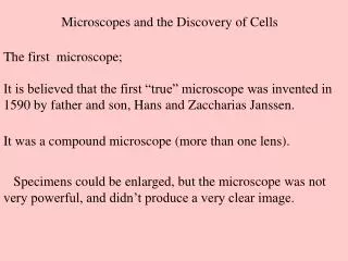
Antony vanLeeuwenhoek Father of microbiology Eyeglass maker who invented first microscope in 1600’s
Eyepiece Body tube Revolving nosepiece Arm Objective lens Stage Stage clips Coarse adjustment knob Fine adjustment Diaphragm Light source Base
Light Microscopy Advantages Can view living specimens Inexpensive and easy to use
Light Microscopy Disadvantages Resolution is limited. Resolution: the ability to form separate images of objects that are close together Resolving power: the minimum distance two points can be separated and still be individually distinguished as two separate points. The smaller the resolving power, the better the resolution.
Light Microscopy Disadvantages • Can only magnify a limited number of times (ours go up to 1000x; best light microscopes magnify up to 4000x) • Limited by focal length of lens
Electron Microscopes To magnify an image a large number of times, you must use an electron microscope. Specimen has a beam of electrons passed through it
Electron Microscopes • There are different types of electron microscopes • In a transmission electron microscope (TEM), an electron beam passes through a very thin section of material • An image is formed because the electrons pass through some parts of the section but not others • In a scanning electron microscope (SEM), a narrow beam of electrons is scanned in a series of lines across the surface of the specimen • The electrons that are reflected or emitted from the surface are collected by a detector and converted into an electrical signal, which is used to build a 3-D image, line by line, on a TV screen
Electron Microscope Advantages Images can be magnified thousands of times (up to 250,000x) A lot of detail can be seen
Electron Microscopy Advantages • Can magnify 1000’s of times • Details are easily visible HIV, magnified 24,000x
Electron Microscopy Disadvantages • Expensive ($$$$$) • Must use heavy metal dyes, which kill organisms
Transmission vs. Scanning EM • Transmission EM’s view cross-sections • SEM’s view surfaces only
To calculate total magnification: • Multiply the magnification on the objective by the magnification found on the eyepiece • You will need this for every specimen you draw under the scope!
Field of View (FOV) • Field of View: Sometimes abbreviated "FOV", it is the diameter of the circle of light that you see when looking into a microscope. • As the power gets greater, the field of view gets smaller. • You can measure this by placing a clear metric ruler on the stage and counting the millimeters from one side to the other. Typically you will see about 4.5mm at 40X, 1.8mm at 100X, 0.45mm at 400X and 0.18mm at 1000X. http://www.microscope-microscope.org/basic/microscope-glossary.htm
Calculating FOV • Measuring the microscope field of view on lowest power • Place a clear plastic ruler with mm markings on top of the stage of your microscope. • Looking through the lowest power objective, focus your image. • Count how many divisions of the ruler fit across the diameter of the field of view. • Multiply the number of divisions by 1000 to obtain the field of view in micrometers (µm). • Record this in µm (1mm = 1000 µm ). • Magnified at 40X, the lines of the ruler are clearly visible. http://www.saskschools.ca/curr_content/biology20/unit1/UNIT1MODULE2LESSON2.htm
FOV Mathematical Calculation Total Magnification Low Power = FOV at Other Power Total Magnification at Other Power FOV at Low Power
Practice calculating FOV Example: • If a 5x FOV is 3 mm, what is the 40x FOV of that microscope? Total Magnification Low Power = FOV at Other Power Total Magnification at Other Power FOV at Low Power 5 = FOV at Other Power 40 3mm (3)(5) = (FOV of higher power)(40) =0.375 mm FOV of higher power
Website for microscope calculations • Calculating with microscopes