Download
1 / 9
90 likes | 243 Views
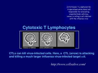
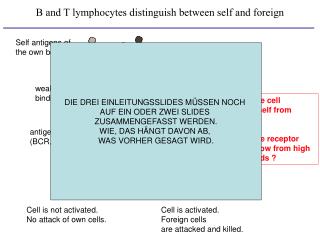
foreign antigen. strong binding. How does the cell distinguish self from foreign ? How does the receptor distinguish low from high affinity ligands ?. Cell is activated. Foreign cells are attacked and killed. B and T lymphocytes distinguish between self and foreign.

E N D
foreign antigen strong binding How does the cell distinguish self from foreign ? How does the receptor distinguish low from high affinity ligands ? Cell is activated. Foreign cells are attacked and killed. B and T lymphocytes distinguish between self and foreign Self antigens of the own body DIE DREI EINLEITUNGSSLIDES MÜSSEN NOCH AUF EIN ODER ZWEI SLIDES ZUSAMMENGEFASST WERDEN. WIE, DAS HÄNGT DAVON AB, WAS VORHER GESAGT WIRD. weak binding antigen receptor (BCR, TCR) T cell Cell is not activated. No attack of own cells.
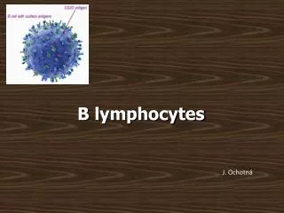
Antigen receptors measure the receptor-ligand affinity independent of the concentration of the ligand. B and T lymphocytes distinguish between self and foreign antigens B cell antigen receptor BCR affinity immune response foreign: self: no response
Model building Old model: New model: 1. 2. 3. receptor-receptor affinity (pre-clustering) Bivalent ligand binding Intracellualr signalling network
Experimental Data: Oligomeric organization of the BCR: BN-PAGE 1 2 Yang and Reth (2010), Nature 1: surface BCR 2: intracellular BCR WB: anti-BCR KA + KD internalization externalization + Formation of a pre-clustered antigen receptor (BCR) Distribution of oligomeric BCR: Mathematical Model: -> dynamic self-association of the receptors
Ligand-binding to pre-clustered antigen receptors (TCR) K1: ligand binding from solution K2: TCR-TCR interaction K3: multimeric ligand binding K2 = 10 (pre-clustered) K2 = 0.1 (clustered on dimer binding) 1/K1=1 mM Bound TCRs Crosslinked TCR pairs -> Receptor pre-clustering increases sensitivity Log [pMHC-dimer], mM Log [pMHC-dimer], mM in cooperation with AG Höfer, Viroquant
self antigens of the own body foreign antigen low affinity high affinity TCR T cell is not activated. No immune response. T cell is activated. Immune response against foreign. T cells can distinguish different affinity ligands lig binding calcium influx Low affinity ligands do not induce Ca flux even at high TCR occupancy. -> T cells can distinguish high from low affinity ligands, largely independent of the ligand concentration.How is this done ?
Ca2+ Ca2+ Ca2+ Ca2+ Different affinity ligand-binding to the TCR total amount of bound antigens high affinity total binding dimeric binding K1=0.1 mM bivalently bound antigens K3=10 intermediate affinity -> Bivalent binding can explain ligand discrimination by T cells: K1=1 mM High affinity ligands bind bivalently and stimulate the TCR. Low affinity ligands bind monovalently and do not stimulate the TCR. K3=1 low affinity K1=10 mM K3=0.1 in cooperation with AG Höfer, Viroquant, & SYBILLA EU FP7
CD19 CD45 mIg CD22 CD72 PIR-B FcgRIIb BTLA CD40 pCD22 DOK3 Gab1 BCAP PAG CSK TRAF3 pCD19 Lyn TRAF2/6 Iga/b SHP-1 Syk PKC CARMA Bcl10 Malt-1 Vav SLP-65 Tab-2 PIP2 Btk HPK-1 PIP2 PIP3 Tak1 IKKg PI3K PKD NIK PTEN PDK Grb-2 PLCg2 DAG IKKb IKKa IP3 SHIP SOS Proteasome Akt Ras RasGRP IkBab p100 Ca2+ Gsk3b Raf activation CaM MEK RelA p50 c-Rel RelB p52 inhibition Calcineurin AND MKP-1 ERK RelA p50 c-Rel RelB p52 RelA RelA RelA feedback NFAT Elk-1 (based on Boolean algebra) Logical model of BCR signalling in cooperation with AG Haus, MaCS
Logical Model of BCR and TCR signalling ► Comparison of BCR signalling network and TCR signalling network: in cooperation with AG Schraven, MaCS Simulation of logical models will help to identify critical elements involved in certain output patterns. -> design of drugs to interfere with the network: treatment of auto-immune diseases and transplant rejections -> improve anti-tumoral immune responses