Download

1 / 92
990 likes | 1.21k Views
SPLEEN. ROY GEORGE SHARON. DEVELOPMENT. Collection of mesenchymal cells in the dorsal mesogastrium- 5 th week Differentiate into lymphoblasts and other blood forming cells Mass projects to left, covered by peritoneum Dorsal mesogastrium gastrosplenic lig.

E N D
SPLEEN ROY GEORGE SHARON
DEVELOPMENT • Collection of mesenchymal cells in the dorsal mesogastrium- 5th week • Differentiate into lymphoblasts and other blood forming cells • Mass projects to left, covered by peritoneum • Dorsal mesogastriumgastrosplenic lig. lienorenal lig.
Contd. • Fusion of lienorenal lig to post.abdominal wall • Change in orientation of stomach • Spleen lies on left side forms the left boundary of the lesser sac of peritoneum
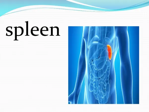
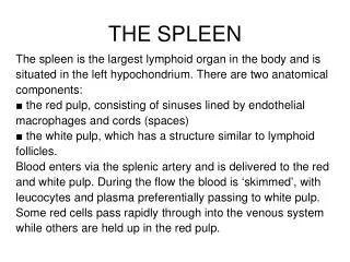
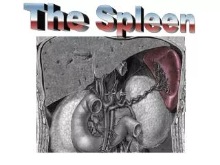
Large encapsulated mass of vascular and lymphoid tissue • SITUATION • Upper Lt. quadrant of the abdomen b/n the fundus of stomach and diaphragm • SHAPE • Slightly curved wedge to ‘domed’ tetrahedron
SIZE • 12cm long , 7cm broad , 3-4 cm wide • WEIGHT • 150 gm (80gm -250gm)
Relations of spleen • SURFACES • Superolateral • Inferomedial • BORDERS • Superior • Inferior • POLES • Anterior • Posterior
SURFACES • DIAPHRAGMATIC • Smooth, convex • Faces superiorly and laterally • related to abd. surface of: • Lt. dome of diaphragm • lower lobe Lt. lung • 9-11 ribs
VISCERAL SURFACE • irregular,faces inferomedially • marked by renal,gastric,pancreatic & colic impressions
Gastric impression • Faces anterolaterally • Broad and concave • Sep. from stomach by peritoneal recess
Renal impression • Concave • Sep. from gastric impression by a raised strip of splenic tissue • Related to: ant. Surface of Lt. kidney superior pole of Lt. suprarenal gland
Colic impression • Inferior pole of spleen • Rel. to splenic flexure of colon & phrenicocolic lig.
Pancreatic impression • Lies b/n colic impression and lateral part of hilum • Rel. to tail of pancreas which lies in the splenorenal lig.
HILUM OF SPLEEN • Lies on visceral surface • A long fissure pierced by several apertures br. of splenic A & V, nerves and lymphatics pass
SUPERIOR BORDER • Sep. diaphragmatic surface from gastric impression • Usually convex • 1-2 notches near the anterior extremity
INFERIOR BORDER • Seperates renal impression from diaphragmatic surface • More blunt and rounded • Corresponds to 11th rib
ANTERIOR POLE • Larger, less angulated • Connects lateral ends of superior & inferior borders • Lies adjacent to splenic flexure and phrenicocolic lig.
POSTERIOR POLE • Angulated • Faces the rounded vertebral column
PERITONEAL CONNECTIONS • Almost entirely covered by peritoneum • Connected to: posterior abd. wallsplenorenal lig. anterolateral abd.wallphrenicocolic lig stomach gastrosplenic lig.
SPLENORENAL LIG • Two layers of peritoneum • Ant. Layer peritoneum of post.wall of lesser sac • Post. Layer -->peritoneum over inf. surface of diaphragm • CONTENTS • Splenic vessels • Tail of pancreas
GASTROSPLENIC LIG. • 2 layers • Ant. Layer from peritoneum lifted of gastric impression • Post. Layer peritoneum over post. aspect of stomach • CONTENTS • Short gastric A. • Lt. gastroepiploic br. of splenic A.
CONGENITAL ANOMALIES • SPLENIC AGENESIS Rare 10% children with heart d/s • POLYSPLENIA Failure of splenic fusion
ACCESSORY SPLEEN(SPLENICULI) Single/multiple 10-30% of population • SITES 50% - Hilum 30% - splenic vessels/behind tail of pancreas remainder – mesocolon/splenic ligaments
NON PARASITIC SPLENIC CYST - rare • TRUE CYST -From embryonal rests 1. dermoid cyst 2.mesenchymal inclusion cyst • FALSE CYST – Trauma - severe pancreatitis - contain serous & haemorrhagic fluid
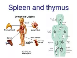
SPLENIC MICROSTRUCTURE • Fibrous framework • White pulp • Red pulp
FIBROUS FRAMEWORK • Tissue capsule • Trabeculae
CAPSULE • Continuous layer, 1.5mm thick • abundant type 1 collagen & elastin • TRABECULAE extend downwards into substance of spleen branch within it supporting framework • Largest enter the hilum • Provide a conduit for splenic A&V,n
WHITE PULP • 0.25-1mm in diameter • White semi- opaque dots on cut section • Aggregations of T lymphocytes terminations of splenic arteriolar adventitia periarteriolar lymphatic sheath ( PALS )
PALS enlarged by aggregation of B lymphocytes LYMPHOID FOLLICLES (MALPIGHIAN BODIES) • Centres of lymphocyte proliferation and differentiation antigenically stimulated GERMINAL CENTRES • Within follicles, splenic arterioles branch to form a series of parallel terminal arterioles PENICILLI
RED PULP • 75% of total splenic volume • Contain VENOUS SINUSES splenic veins • Sep. by fibrocellular network RETICULUM fibroblasts (reticular cells) & collagen(splenic macrophages)
VENOUS SINUSES • Elongated ovoid vessels 50microm. • Lined by incomplete endothelium aligned along long axis (stave cells) attached by intercellular junctions (blood passes from splenic cords to sinus lumen) luminal &external surface bears short irregular microvilli Supported circumferential &longitudinal reticular fibres
MARGINAL ZONE • At interface of red pulp and white pulp • Region where the blood is delivered to the red pulp • Lymphocytes leave the circulation to migrate to T and B lymphocyte areas of the white pulp • Aggregation of macrophages surround arteriolar ends- periarteriolar macrophage sheath (Schweigger-Seidel)
SURFACE ANATOMY • Spleen is marked on Lt. side of back with long axis along line of 10th rib • Upper border upper border of 9th rib • Lower border lower border of 11th rib • Ant. extends to mid –axillary line • Post. 4cm lateral to 10th thoracic spine
VASCULAR SUPPLY • ARTERIAL SUPPLY • Splenic A. – large,tortuous • Divides in lienorenal lig. into 2-3 main branches4-5 segmental br. • In each segment ,ramifies within trabeculae to supply parenchyma and capsule
VENOUS SUPPLY • Minor veins pass from red pulp segmental veinsdrain into main splenic V. in lienorenal lig. • LYMPHATIC SUPPLY • Drain along trabeculae • Pancreaticosplenic and coeliac nodes
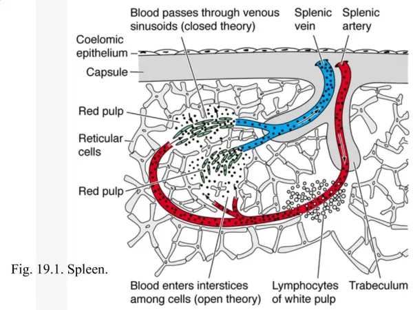
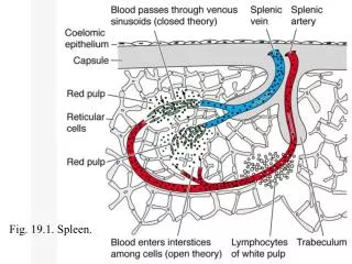
SPLENIC MICROCIRCULATION • OPEN CIRCULATION • Arteriolar blood reticular spaces in splenic cords enter sinuses through minute slits • CLOSED CIRCULATION • Arteriolar blood venous sinuses
NERVE SUPPLY • Arise from coeliac sympathetic plexus • Mainly adrenergic vasomotor • Regulate of blood flow through spleen
PHYSIOLOGY • As plasma rich blood pass through sinuses: 1.filtered 2.particles phagocytosed sinus splenic pulp pressure reflects pressure throughout the portal system90% blood course through OPEN circulation
CONTD. • ERYTHROCYTE CLEARANCE • Abnormal, aged cleared • Approx. 20 ml red cells removed/day • Factors: • change in biophysical properties • hypoxic, acidotic, glucose deprived environment
fall in cellular ATP levels • loss of ATP dependent functions • sodium and calcium efflux • loss of membrane integrity