Download
1 / 24
250 likes | 622 Views
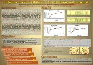
Mechanotransduction, Tensegrity and Durotaxis. ChemEng 575: Lecture 14 April 8 th , 2014 Reading: 3 Papers online. In Lecture 8. We discussed ways to test and quantify the mechanical properties of materials

E N D
Mechanotransduction, Tensegrityand Durotaxis ChemEng 575: Lecture 14 April 8th, 2014 Reading: 3 Papers online
In Lecture 8 • We discussed ways to test and quantify the mechanical properties of materials • Left you with food for thought: is that important for tissue engineering design? (and your grant)? • Question for today: do cells care? • i.e. can cells sense and respond to mechanical forces?
Mechanotransduction • The ability of a cell to turn a mechanical cue from the ECM into an intracellular signal • RhoA, pSrc, pAkt • Or a phenotypic response • Migration, differentiation, shape, growth
Where might mechanotransduction be important in your body? • Class poll: where are cells exposed to mechanical forces?
Mechanotransduction: Cell can translate Mechanical Information from the ECM to an intracellular biochemical signal “Mechanotransduction”
How does this happen? • Focal adhesions. • Remember, those connections between integrins and the actin cytoskeleton in a cell. • When, how do focal adhesions re-arrange in response to mechanical forces? S S P S=structural P=signaler S P S S S P
Two different ways this can happen OUTSIDE-IN (ECM-initiated) INSIDE-OUT (cell-initiated)
Vibrating Cells (outside in signaling)Cells will pull at the site of vibration Go to http://www.plosone.org/article/info%3Adoi%2F10.1371%2Fjournal.pone.0026181#s5 Nishitani, PLOS 1
Pulling on cell attachment points (Outside-In)Focal adhesions are recruited to the site of stretch
Stretching the underneath substrate (Outside-In)Microtubules assemble (polymerize) when cell is stretched Putnam et al., JCS, 1998
Proposed: Cell-ECM force balance through F-actin and microtubules Courtesy of A. Putnam • In response to extracellular stretch or an intrinsic ECM stiffness, F-actin microfilaments adjust in tensional resistance, and the microtubule network adjusts in compressive resistance.
Tensegrity: a Physical Mechanism of Mechanotransduction Cytoskeleton connects from focal adhesions to nucleus. Forces at focal adhesions can propogate to changes in shape of nucleus affects transcription regulators gene expression/phenotype
Traction Force Microscopy: Tool to Measure Cellular Forces Exerted on Substrate
Phenotypic result • Either because of signaling changes at the site of focal adhesions… • Or through this force balance which eventually stretches the nuclear membrane… • Stiffness of the ECM can regulate: • Stem cell differentiation (bone v nerve v muscle) • Cell growth (many cell types) • Cell migration
Polyacrylamide as a Biomaterial Varying acrylamide and bis-acrylamide Polyacrylamide disc Heterobifunctionalcrosslinkersulfo-SANPAH Fibronectin Peyton, S.R. and Putnam, A.J. J. Cell. Phys. 2005 Jul;204(1):198-209.
1: Step Changes in Stiffness 3T3 Fibroblasts on PAA Migrate from soft-to-stiff substrates Biophys J. Lo et al. (2000) 79;144-152
Durotaxis: gradientsvia photomask polymerization Wong, J. Langmuir, 2003
Durokinesis: Biphasic Migration Dependence on Substrate Stiffness Speed (um/hr) • Durokinesis: SMCs migrate fastest on an ‘optimally stiff’ substrate • Lecture 9: actin polymerization controlled by adhesive protein density as well (Haptokinesis). • Cells need stiffer substrate when less fibronectin is attached to surface to migrate at maximum capacity Substrate stiffness Peyton and Putnam, J. Cell. Phys. 2005
Cytoskeletal Assembly Regulated by Substrate Stiffness Peyton and Putnam, J. Cell. Phys. 2005
Biomaterials to Study Durotaxis/Durokinesis • Synthetic Polymers • Polyacrylamide (PAA), Poly(ethylene glycol) (PEG), Polydimethylsiloxane (PDMS) • Independent tunability • Wide range of mechanical properties (100Pa – MPa) • Difficult chemistries • Not always 3D transferable • Natural Biopolymers • Collagen, Fibrin, Matrigel • Contain cell-adhesive domains, 3D transferable • Natural chemistries • Soft 1Pa-10kPa • Lumped parameters • In Vivo Tissue Elastic Moduli Range • Brain: 100s of Pa • Liver: 10-100 kPa • Artery: ~40kPa • Skin: ~100 kPa • Bone: 100s of MPa to GPa
3D Collagen: Results Influenced by Polymerization Conditions Native bovine dermal type I collagen Motility requires MT1-MMP (Nutragen) JCB Wolf et. al. (2003) Native bovine dermal type I collagen Motility can be protease-independent (Vitrogen, pepsin-extracted, non-covalent crosslinks) MBC Kim et. al. (2008) Freeze-dried Collagen-GAG 1D migration along fibers Biophys J Harley et. al. (2008)
Cell-Secreted ECMs: 3D = 1D? JCB Doyle et. al. (2009)