Download
1 / 48
490 likes | 595 Views
Explore the fascinating world of protein functions and synthesis, from primary to quaternary structures, evolutionary advantages, genetic coding, and transcription processes in prokaryotes and eukaryotes.

E N D
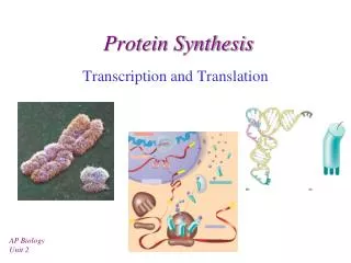
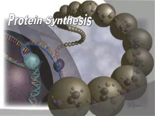
Protein Synthesis and Structure Section 2-4
Protein Functions: General Information • Proteins account for almost 50% of the dry mass of most cells • Proteins are the most structurally sophisticated molecules known • Each protein has a specific 3-diminsional shape, or “confirmation” that is vital to its function
Protein Functions • Enzymatic • Structural • Storage • Transport • Hormonal • Receptor • Contractile and motor • Defensive
Primary Structure • The unique sequence of amino acids that make up a polypeptide • Amino acids: • An asymmetrical carbon • An amino group (contains Nitrogen) • A carboxyl group • A side chain (R group) which makes each amino acid unique
Primary Structure • Amino acids are linked together with dehydration reactions • Peptide bonds- the bonds between amino acids
Primary Structure • The chain will have two ends: • An amino end- known as the N-terminus • A carboxyl end- known as the C-terminus • Primary structure is achieved through the processes of transcription and translation
Secondary Structure • Refers to the coils and folds in the polypeptide chain due to hydrogen bonds between repeating areas on the polypeptide backbone
Secondary Structure • Alpha helix (α helix)- a delicate coil held together by hydrogen bonding between every fourth amino acid • Beta pleated sheet (β pleated sheet)- two or more regions of a polypeptide chain lying side by side and connected by hydrogen bonds between the two parallel polypeptide back bones
Tertiary Structure • Folding due to interactions between the amino acid side chains • Main causes of tertiary structure • Hydrogen bonds between polar R-groups • Hydrophobic interactions between nonpolar R groups causes them to clump together and form Van der Wall’s interactions • Dislufide bridges between two sulfhydryl groups
Tertiary Structure: Main Causes • Hydrogen bonds between polar R-groups • Hydrophobic interactions between nonpolar R groups causes them to clump together and form Van der Wall’s interactions • Dislufide bridges between two sulfhydryl groups
Quaternary Structure • Overall shape of the protein caused by the association of two or more polypeptide chains • NOT ALL PROTEINS HAVE QUATERNARY STRUCTURE
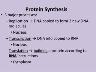
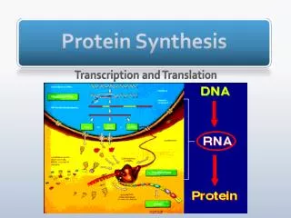
General Information • Also called gene expression • DNA provides the blueprints for the building of proteins
General Information • Involves two processes: • Transcription- copying DNA into mRNA • Translation- translates the code from nucleic acid into amino acid at the ribosome
Evolutionary Advantage of Transcription and Translation • DNA is protected inside the nucleus • Using an RNA intermediate allows multiple copies of a protein to be made at once because many mRNA molecules can be made from one gene, then translated repeatedly
Prokaryotes vs. Eukaryotes Prokaryotes • Only one compartment (no nucleus) • Transcription and Translation occur simultaneously
Prokaryotes vs. Eukaryotes Eukaryotes • Transcription occurs in the nucleus • The primary transcript is then modified (RNA processing) before leaving the nucleus • Translation occurs in the cytoplasm at the ribosome
The Genetic Code • Triplet Code- the flow if information from gene (DNA) to protein is written in the DNA as non-overlapping, three-nucleotide segments • Template Strand: • The mRNA is complimentary to the template strand • The DNA is read in the 3’ to 5’ • The mRNA is synthesized and read from 5’ to 3’
The Genetic Code • Codons- each three base sequence on the mRNA strand • Each codon codes for a specific amino acid
Redundant but not Ambiguous • Redundant- multiple codons can code for the same amino acid • Not Ambiguous- no codon codes for more than one amino acid
Special Codons • AUG= start • UAA, UAG, UGA= stop
Initiation • RNA polymerase binds to the promoter • The promoter is a specific sequence that tells the RNA polymerase where to bind and determines what DNA strand will serve as the template • In eukaryotes, specific proteins called transcription factors assist the RNA polymerase in binding and forming the transcription initiation complex
Elongation • RNA polymerase adds nucleotides to the 3’ end of the growing RNA molecule • Complimentary base pairing occurs • The new RNA molecule peals away from the DNA template and the DNA reforms
Termination • In prokaryotes, the RNA polymerase detaches after the termination signal is transcribed • In eukaryotes, the RNA polymerase transcribes the polyadenylation signal sequence then the mRNA is cut off of the RNA polymerase
RNA Processing EUKARYOTIC CELLS ONLY
Altering of the Ends of the mRNA • 5’ cap- modified guanine molecule added on the 5’ end • Poly-A-tail- 50-250 adenine nucleotides are added to the 3’ end • Functions: • Facilitate export from the nucleus • Protect the mRNA from degradation by enzymes • Assist the ribosomes in attaching in the cytoplasm
RNA Splicing • Removal of large portions of the mRNA • snRNPs (“snurps”) recognize and cut out areas of the mRNA • Introns- the portions of the mRNA that are removed • Exons- the portions of the mRNA that exit the nucleus
Transfer RNA, tRNA • Translates nucleotides into amino acids • One end has an anticodon, complementary to the mRNA codon • The other end is bound to an amino acid • Excellent example of how structure fits function
Ribosomes • Contain three sites for holding tRNA: • P site- holds the growing polypeptide chain • A site- holds the tRNA that is carrying the next amino acid in the chain • E site- where the tRNA leaves the ribosome • Exit Tunnel= where the polypeptide leaves the ribosome
Initiation • Small ribosomal subunit binds the mRNA and the initiator tRNA • Subunit scans the mRNA until it reaches the start codon, establishing the correct reading frame as the tRNA hydrogen bonds to the start codon
Initiation • Translation initiation complex forms- the large ribosomal subunit attaches with the assistance of initiation factors and an expenditure of energy
Elongation • The ribosome reads the mRNA in the 5’ to 3’ direction • Anticodon of the incoming tRNA hydrogen bonds to the mRNA codon in the A site • The peptide bond forms between the amino acid on the tRNA of the A site and the growing polypeptide chain in the P site • Translocation of the tRNA shifts the A site tRNA to the P site and the P site tRNA to the E site so it can exit
Termination • Release factor: • Added when stop codon is reached • Causes the addition of a water molecule to the end of the polypeptide • The polypeptide is released
Protein Folding • Folding occurs as the protein is being synthesized • Folding is dependent on • The properties of the peptide chain • The physical and chemical properties of the environment WHY MIGHT THIS BE A PROBLEM???
Chaperonins • Proteins that assist in the proper folding of other proteins by shielding them form the cell environment
Post-Translational Modification • Chemical modification by the attachment of sugars, lipids, phosphate groups, or other components • Enzymes may remove one or more amino acids from the N-terminus • Single polypeptides may by cut into two or more smaller pieces
Denaturation • The changes in a protein’s native conformation that renders it biologically inactive • Factors that cause denaturation: • Change in the environment • Change in temperature • Change in pH
Changes in Environment • If moved from an aqueous environment to a nonpolar organic solvent, the protein will turn inside out • Chemicals can disrupt disulfide and hydrogen bonds that stabilize secondary and tertiary structure
Changes in Temperature • Excessive heat can cause movement to overpower sensitive hydrogen bonds • Excessive cold will slow the protein down substantially
Changes in pH • All proteins have an optimal pH at which they function • Optimal pH is not necessarily 7