Download
1 / 45
480 likes | 607 Views
Anatomy of the Heart. Video. Heart Shape and Make-up . Nine (9) inches long x three (3) inches wide. Size of a fist Triangular Involuntary striated muscle tissue Only found in this organ. Beats 60-100 times per minute 2.5 billion times during an average lifespan. .

E N D
Anatomy of the Heart Video
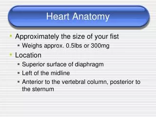
Heart Shapeand Make-up • Nine (9) inches long x three (3) inches wide. • Size of a fist • Triangular • Involuntary striated muscle tissue • Only found in this organ. • Beats 60-100 times per minute • 2.5 billion times during an average lifespan.
Heart Location • Base--attached to several large blood vessels and lies beneath the second rib. • Apex— • at the fifth intercostal space • Points towards the left • Located within the mediastinum, bordered laterally by the lungs, posterior is the backbone, and anterior is the sternum
Pericardial Cavity • Pericardium • Located between the parietal and visceral layers • Fluid filled sack that surrounds the heart • Pericardial fluid • Several functions • Keeps the heart contained in the chest cavity • Prevents the heart from over expanding when blood volume increases • Prevents friction (rubbing with beats)
Layers • Made up of three (3) distinct layers: • Outer epicardium • Myocardium • Inner endocardium
Epicardium • Corresponds to the visceral pericardium. • Functions as an outer protective layer. • Serous membrane that consists of connective tissue covered by epithelium. • Includes blood capillaries, lymph capillaries, and nerve fibers.
Myocardium • Relatively thick. • Consists largely of cardiac muscle tissue responsible for forcing blood out of the heart chambers. • Muscle fibers are arranged in planes, separated by connective tissues that are richly supplied with blood capillaries, and nerve fibers.
Endocardium • Consists of epithelial and connective tissue that contains many elastic and collagenous fibers. • Connective tissue also contains blood vessels and some specialized cardiacmuscle fibers called Purkinje fibers. • Lines all of the heart chambers and covers heart valves. • Is continuous with the inner lining of blood vessels--endothelium.
READ Handout
Anatomy of the Heart Tuesday
Vena Cava • Superior Vena Cava • Bringing de-oxygenated blood • Upper body to the right atrium of the heart • Inferior Vena Cava • Bringing de-oxygenated blood • Lower body to the right atrium of the heart
Chambers • Atria • Two chambers at the top • Right • Receives deoxygenated blood from the superior and inferior vena cava • Left • It receives oxygenated blood from the pulmonary veins
Chambers • Ventricles • Two chambers at the bottom • Right • It receives deoxygenated blood from the right atrium • Left • It receives oxygen richblood from the left atrium • Video
Coronary Arteries • Coronary arteries supply blood to the heart muscle • oxygen-rich blood to function • oxygen-depleted blood must be carried away • consist of two main arteries • the right and left coronary arteries.
Valves • Tricuspid • valve is between the right atrium and right ventricle. • Pulmonary • valve is between the right ventricle and the pulmonary artery.
Valves • Mitral (Bicuspid) • valve is between the left atrium and left ventricle. • Aortic • valve is between the left ventricle and the aorta. • Each valve has a set of flaps (also called leaflets or cusps). When working properly, the heart valves open and close fully.
Vessel Valves • Pulmonary Valve • semilunar valve of the heart that lies between the right ventricle and the pulmonary artery and has three cusps. • Aortic Valve • It lies between the left ventricle and the aorta • video
READ Handout
Anatomy of the Heart Wednesday
Importance of Coronary Arteries • Coronary artery disorder or disease • Reducing the flow of oxygen and nutrients to the heart • leads to a heart attack and possibly death. • Video • Atherosclerosis (a build-up of plaque in the inner lining of an artery causing it to narrow or become blocked) is the most common cause of heart disease.
Vessels • Video • Superior Vena Cava • Inferior Vena Cava • Aorta • Pulmonary Artery • Pulmonary Vein
Aorta • Largest single blood vessel in the body • diameter of your thumb • carries oxygen-rich blood from the left ventricle to the various parts of the body • Aortic Arch • Top curve of the aorta
READ Handout
Anatomy of the Heart Thursday
Pulmonary Artery • Vessel transporting de-oxygenated blood from the right ventricle to the lungs • A common misconception is that all arteries carry oxygen-rich blood • It is more appropriate to classify arteries as vessels carrying blood away from the heart.
Pulmonary Vein Vessel transporting oxygen-rich blood from the lungs to the left atrium A common misconception is that all veinscarry de-oxygenated blood it is more appropriate to classify veins as vessels carrying blood to the heart Video-blood flow Video
Entire Cycle • video- blood flow • Lyrics
READ Handout