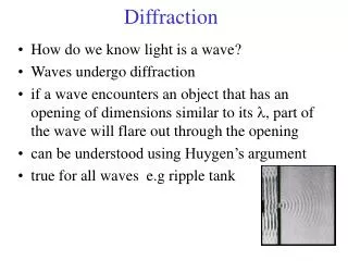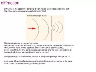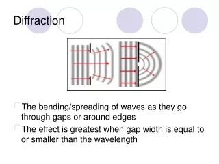
Diffraction
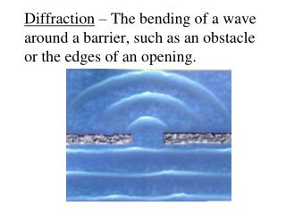
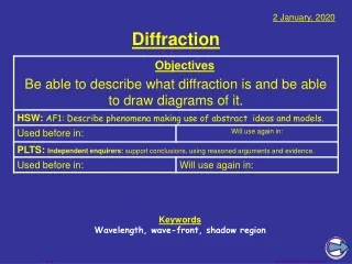
Diffraction. When “s cattering” is not random. scattering. detector. detector. x-ray beam. sample. Scattering: atom by atom. intensity. h index. Scattering: atom by atom. intensity. h index. θ. d ∙ sin( θ ). Bragg’s Law. to detector. n λ = 2d sin( θ ). atom #1. d.

Diffraction
E N D
Presentation Transcript
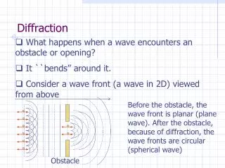
Diffraction When “scattering” is not random
scattering detector detector x-ray beam sample
Scattering: atom by atom intensity h index
Scattering: atom by atom intensity h index
θ d∙sin(θ) Bragg’s Law to detector nλ = 2d sin(θ) atom #1 d to source atom #2
scattering from a lattice colored by phase sample detector
scattering from a molecule colored by phase sample detector
scattering from a crystal structure colored by phase sample detector
Spot shape Ewald sphere Φcircle (h,k,l) λ* d* spindle axis diffracted ray λ*
mosaic spread Ewald sphere Φcircle (h,k,l) d* spindle axis diffracted rays d*
Ewald sphere beam divergence Φcircle (h,k,l) d* λ* spindle axis diffracted ray λ*
spectral dispersion Ewald sphere Φcircle (h,k,l) λ’* d* spindle axis diffracted ray λ’*
spot shape Ewald sphere Ewald sphere Ewald sphere Ewald sphere Φcircle Φcircle Φcircle Φcircle (h,k,l) (h,k,l) (h,k,l) (h,k,l) λ* λ’* d* d* d* d* λ* spindle axis spindle axis spindle axis spindle axis diffracted rays d* diffracted ray diffracted ray diffracted ray λ’* λ* λ*
10 Å Now What?
Resolution http://bl831.als.lbl.gov/~jamesh/movies/resolution.mpeg
B-factor What is “disorder”? order order disorder
“B” factors ATOM 122 N LEU A 13 -3.244 25.808 19.998 1.00 16.96 N ATOM 123 CA LEU A 13 -2.877 25.448 21.355 1.00 15.29 C ATOM 124 C LEU A 13 -2.792 23.966 21.561 1.00 17.54 C ATOM 125 O LEU A 13 -1.814 23.493 22.143 1.00 16.35 O ATOM 126 CB LEU A 13 -3.907 26.164 22.268 1.00 18.72 C ATOM 127 CG LEU A 13 -3.577 25.982 23.738 1.00 21.19 C ATOM 128 CD1 LEU A 13 -2.283 26.820 24.019 1.00 19.43 C ATOM 129 CD2 LEU A 13 -4.702 26.474 24.639 1.00 24.65 C ATOM 130 N SER A 14 -3.677 23.149 20.979 1.00 15.96 N ATOM 131 CA SER A 14 -3.646 21.711 21.061 1.00 18.26 C ATOM 132 C SER A 14 -2.373 21.203 20.360 1.00 18.71 C ATOM 133 O SER A 14 -1.747 20.315 20.930 1.00 17.47 O ATOM 134 CB SER A 14 -4.875 21.077 20.419 1.00 17.62 C ATOM 135 OG ASER A 14 -4.825 19.665 20.388 0.50 20.89 O ATOM 136 OG BSER A 14 -6.027 21.408 21.164 0.50 18.67 O ATOM 137 N LYS A 15 -2.045 21.772 19.215 1.00 18.03 N ATOM 138 CA LYS A 15 -0.799 21.361 18.555 1.00 18.12 C ATOM 139 C LYS A 15 0.446 21.707 19.351 1.00 18.81 C ATOM 140 O LYS A 15 1.400 20.948 19.411 1.00 17.77 O ATOM 141 CB LYS A 15 -0.700 22.034 17.177 1.00 14.49 C ATOM 142 CG LYS A 15 -1.727 21.368 16.256 1.00 16.12 C ATOM 143 CD LYS A 15 -1.663 22.147 14.936 1.00 19.40 C ATOM 144 CE ALYS A 15 -2.725 21.614 13.986 0.50 17.42 C ATOM 145 CE BLYS A 15 -1.750 21.211 13.750 0.50 17.01 C ATOM 146 NZ ALYS A 15 -2.346 21.674 12.559 0.50 18.61 N ATOM 147 NZ BLYS A 15 -3.052 20.513 13.741 0.50 18.76 N
“B” factors B = 8π2 ux2 ux= RMS variation perpendicular to plane
“B” factors electron density (e-/Å3) position (Å)
“B” factors B ≈ 4d2 + 12 d = resolution in Å essentially, the “resolution” of an atom
Debye-Waller-Ott factor F = F0 exp( - A∙s - B∙s2 - C∙s3 - … ) F - structure factor A - something Debye said was zero B - canonical Debye-Waller factor C - something else Debye said was zero s - 0.5/d d - resolution of spot (Å)
Gaussian Exponential Debye-Waller-Ott factor Reciprocal Space normalized total intensity 5 2.5 1.7 1.25 1.0 Resolution (Ǻ)
Gaussian Lorentzian Debye-Waller-Ott factor Direct Space normalized number of atoms magnitude of displacement (Å)
Wilson plot Rcryst/Rfree 0.355/0.514 0.257/0.449 0.209/0.407 scaled <F2> 4.1 3.5 3.2 2.9 2.7 2.5 2.4 2.2 2.1 resolution (Å) (sin(θ)/λ)2
Purity is crucial! important for resolution McPherson, A., Malkin, A. J., Kuznetsov, Y. G. & Plomp, M. (2001)."Atomic force microscopy applications in macromolecular crystallography", Acta Cryst. D57, 1053-1060. not important for initial hits
Purity! is 95% good enough? 99%? Purity! conformational (homogeneous) Purity! kinetic (stable over time) What can I improve?
add a column fractional recrystallization heat shock mutate Lys avoid stress What can I improve? Newman J. (2006) Acta Cryst. D62 27-31.
causes of stress physical contact don’t touch the part you intend to shoot osmotic shock equilibrate, or calculate matching solution changes in dielectric constant Petsko (1975) J. Mol. Biol.96, 381-388. cooled density mismatch Juers & Matthews (2004) Acta Cryst. D60, 412-421. basically: no sudden moves!
Completeness: missing wedge http://bl831.als.lbl.gov/~jamesh/movies/osc.mpeg
Non-isomorphism in lysozyme RH 84.2% vs71.9% • Riso = 44.5% RMSD = 0.18 Å
“photon counting” Read-out noise Shutter jitter Beam flicker spot shape radiation damage σ(N) = sqrt(N) rms 11.5 e-/pixel rms 0.57 ms 0.15 %/√Hz pixels? mosaicity? B/Gray? signalvsnoise
fractional noise “photon counting” constant noise σ(I) = k x I “% error” σ(I) = k x sqrt(I) σ(I) = k signalvsnoise
adjust exposure so this is ~100 Optimal exposure time(faint spots) thr Optimal exposure time for data set (s) tref exposure time of reference image (s) bgref background level near weak spots on reference image (ADU) bg0 ADC offset of detector (ADU) bghr optimal background level (via thr) σ0rms read-out noise (ADU) gain ADU/photon m multiplicity of data set (including partials)
anomalous scattering x-ray beam sample detector
anomalous signal √ ΔF F # sites MW (Da) ≈1.2 f” World record! ΔF/F = 0.5% Wang, Dauter & Dauter (2006) Acta Cryst. D62, 1475-1483. Crick, F. H. C. & Magdoff, B. S. (1956) Acta Crystallogr.9, 901-908. Hendrickson, W. A. & Teeter, M. M. (1981) Nature290, 107-113.
no “scale factor” is perfectly known • no source of light is perfectly stable • no shutter is perfectly reproducible • no crystal is perfectly still • no detector is perfectly calibrated Fractional error
Vxtal λ3 L Vcell ωVcell Darwin’s Formula I(hkl) = Ibeam re2 P A | F(hkl) |2 I(hkl) - photons/spot (fully-recorded) Ibeam - incident (photons/s/m2 ) re - classical electron radius (2.818x10⁻¹⁵ m) Vxtal - volume of crystal (in m3) Vcell - volume of unit cell (in m3) λ - x-ray wavelength (in meters!) ω - rotation speed (radians/s) L - Lorentz factor (speed/speed) P - polarization factor (1+cos2(2θ) -Pfac∙cos(2Φ)sin2(2θ))/2 A - attenuation factor exp(-μxtal∙lpath) F(hkl) - structure amplitude (electrons) C. G. Darwin (1914)
tso txo tsi txi tso txo txi tsi tso txo IT Ibeam txi tsi attenuation factor μsolvent μxtal A = = exp[-μxtal(txi+ txo) -μsolvent(tsi + tso)] Bouguer, P. (1729). Essai d'optique sur la gradation de la lumière. Lambert, J. H. (1760). Photometria: sive De mensura et gradibus luminis, colorum et umbrae. E. Klett. Beer, A. (1852)."Bestimmung der Absorption des rothen Lichts in farbigen Flüssigkeiten", Ann. Phys. Chem86, 78-90.
Ewald sphere Lorentz Factor Φcircle (h,k,l) diffracted ray spindle axis
% error from rad dam Riso≈ 0.7 %/MGy Riso (%) change in dose (MGy) data taken from Banumathi, et al. (2004) Acta Cryst. D60, 1085-1093.
Beam Flicker 1/f noise or “flicker noise” comes from everything
Shutter Jitter open closed shutter jitter
xtal vibration noise diffracted beam incident beam
Shutter Jitter CC to correct model rms timing error (% exposure)
Beam Flicker CC to correct model flicker noise (%/√Hz)
Solution to vibration: attenu-wait! • reduce flux • increase exposure