Download
1 / 31
310 likes | 331 Views
This tutorial provides information and questions to test your knowledge on the back part of the eye. Learn about the structure and function of the posterior chamber, retina, photoreceptors, and more.

E N D
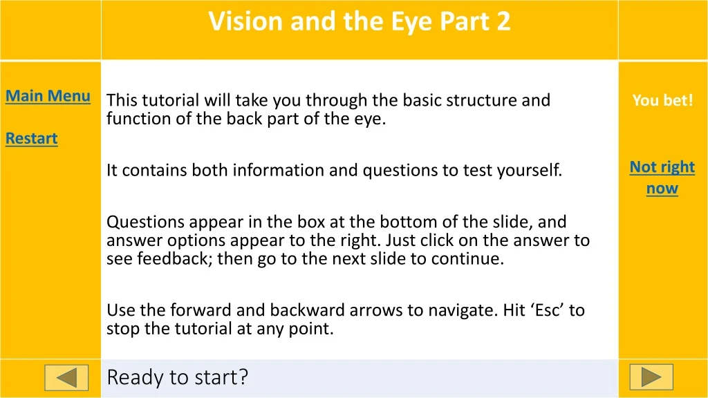
Front Back You bet! • This tutorial will take you through the basic structure and function of the back part of the eye. • It contains both information and questions to test yourself. • Questions appear in the box at the bottom of the slide, and answer options appear to the right. Just click on the answer to see feedback; then go to the next slide to continue. • Use the forward and backward arrows to navigate. Hit ‘Esc’ to stop the tutorial at any point. Not right now Ready to start?
Posterior chamber Good work! You figured it out. Check again! The pupil‘s in the middle of the iris. Does that word really refer to the back ? Pupil Anterior chamber Image modified from Wikipedia, https://en.wikipedia.org/wiki/Mammalian_eye used under a Creative Commons license The part of your eye behind the iris is the _____
That’s right! The sclera is the white of your eye. The cornea’s completely transparent. The iris is inside your eye! Sclera Cornea Iris Image modified from Wikipedia, https://en.wikipedia.org/wiki/Mammalian_eye used under a Creative Commons license The white layer covering the outside of the eye is the:
Retina Right! That’s where the neurons are. The iris is mostly muscle, not neurons. The lens focuses light, but doesn’t detect it. Iris Lens Image modified from Wikipedia, https://en.wikipedia.org/wiki/Mammalian_eye used under a Creative Commons license Light rays are detected when they hit the:
Retinal arteries These are in front of the retina, not behind it! The cornea is the clear layer in the front of the eye Right! Cornea Choroid Image modified from Wikipedia, https://en.wikipedia.org/wiki/Mammalian_eye, used under a Creative Commons license The layer of blood vessels between the sclera and retina is the ________
Aqueous humor Nice try, but this is in the anterior chamber. Good work! Retinal arteries don’t fill the chamber. Vitreous humor Retinal arteries Image modified from Wikipedia, https://en.wikipedia.org/wiki/Mammalian_eye, used under a Creative Commons license The jelly-like substance that fills the posterior chamber, pushing the retina against the choroid, is the _______
Good work on the anatomy! • The posterior chamber of your eye is all about REPORTING light. • The photoreceptors in the retina change their firing in response to light, and transmit that information to the brain. Image modified from Rev. XanatosSatanicosBombasticos (ClintJCL): used under a Creative Commons license
Let’s look at the cells that do this work. • The retinas develop as outpouchings of your brain, so they are actually brain tissue! • This is the brain and retinas of an early chick embryo, but humans develop the same way. Image from Wikipedia, https://en.wikipedia.org/wiki/Eye_development Used under a Creative Commons license
Let’s look at a close-up of the retina. • The photoreceptors are in the posterior side of the retina, right next to the choroid blood vessels so they can get lots of nutrients and Oxygen. photoreceptors choroid
The photoreceptors synapse with the bipolar cells. • The bipolar cells synapse with the ganglion cells. • The ganglion cells send their axons out through the optic nerve, all the way into the brain. Bipolar cell photoreceptors choroid Ganglion cells Optic nerve
Rods are rod-shaped That’s right! An easy way to remember is ‘c for color.’ They are photosensors, but you can be more specific. Rods • The photoreceptors in this picture are named for their shape. • I drew them in different colors to indicate that they respond to light of different colors- red, green, and blue. Bipolar cell photoreceptors choroid Cones Ganglion cells Photo-sensors Optic nerve What’s their name?
They do both detect red light, but in different places! The brain doesn’t know what they are, only if they sent it a message. That’s right! The brain gets a different message from each cell! • Now I’ll take some of them away so you can see more clearly. • Suppose you look at two objects. The light from one focuses and hits cone A, and the light from the other hits cone B. • Can your brain tell which cone the light hit? No, because they both detect red light choroid No, because they’re both cones A B Optic nerve Yes, because they have separate connections to the brain Can your brain distinguish them?
Good! Each cone sends its own message to the brain. Because each cone sends its own message, the dots are distinct Oops - Each cone detects only one color. If light hits all these cones at once, what will your brain detect?
Each of your cones has its very own bipolar cell and ganglion cell – its own hotline to the brain! • So your brain can tell what each individual cone is seeing. That’s how you can distinguish small objects from one another. • That ability is called ACUITY. Your cones give you very ACUTE vision. • So cones give you acute color vision. The down side is – they need a lot of light to work. They are not very SENSITIVE in low light.
The only cells that fire in response to light are photo-receptors. Right! There are no photo-receptors here to detect the light. True – but if light doesn’t hit photo-receptors, your brain will never know about it. • Before we go away from this diagram of the back of your retina, answer another question. • What will happen if light focuses on point C of this retina? A big blur of light, because all the axons are stimulated choroid Nothing, because it won’t hit any photo-receptors A B C Optic nerve White light, because it won’t hit any cones What will the brain detect?
Where the axons exit your retina into the optic nerve, you have a BLIND SPOT – a place where there are no photoreceptors to detect light. When you look around, do you see a big blank spot? That’s right! Your brain fills the blind spot in with whatever is hitting the cones near it. If light hits all these cones at once, what will your brain detect?
Rods • Let’s look at a close-up of a different part of the retina – toward the edge. • Just like before, the photoreceptors are right next to the choroid blood vessels so they can get lots of nutrients and Oxygen. But they look different. That’s right! They are rod-shaped. But you saw cones had a different shape… Can you be more specific? Which kind of photosensor? photoreceptors choroid Cones Photo-sensors What do you think these photoreceptors are called?
Just like cones, rods synapse with bipolar cells. The bipolar cells then synapse with ganglion cells. • But it’s not quite the same, is it? That’s right! Rods just distinguish light and dark. What else? That’s right! Many rods share one bipolar cell. What else? That’s right! Many rods share one ganglion cell. What else? They all see the same color photoreceptors photoreceptors choroid There aren’t enough bipolar cells Bipolar cell Ganglion cell There aren’t enough ganglion cells What’s different?
If light hits cell A on this part of the retina, can your brain tell it apart from light hitting cell B? But the brain only knows what the ganglion cell tells it! Right! No matter which of these rods fires, the same ganglion cell sends a message to the brain. Yes, they’re different cells photoreceptors photoreceptors choroid A No, they use the same axon to the brain B Bipolar cell Ganglion cell What’s different?
If light hits a cell on this part of your retina, what will your brain detect? But the brain only knows what the ganglion cell tells it! Does it know which rod fired? Right! The brain just knows one of them fired, but not which one. photoreceptors photoreceptors choroid A B Bipolar cell Ganglion cell What’s different?
The cones are located in the back of your eye, in the central portion of your retina. The rods are located in the peripheral parts of your retina. • That means cones see what’s right in front of you, while rods see the things that are off to the sides. Image modified from Wikipedia, https://en.wikipedia.org/wiki/Mammalian_eye used under a Creative Commons license
So when you watch the beautiful balloons… • what your rods and cones actually detect is more like this. Image modified from Wikipedia, https://en.wikipedia.org/wiki/Hot_air_balloon_festival used under a Creative Commons license
How do you see the whole scene in color? • You keep moving your eyes back and forth, and your brain remembers the colors you just looked at. It fills them in, the same way it fills in your blind spot. Image modified from Wikipedia, https://en.wikipedia.org/wiki/Hot_air_balloon_festival used under a Creative Commons license
The orange ones Check again. Light from that ‘V’ has to enter the eye through the pupil – where will it hit the retina? Right! The light from the ‘V’ will hit the blue part of the retina on both eyes. • You’re looking down on someone’s eyes and brain. The two sides of the retina and their nerve tracts have been color-coded. The blue ones Image from Wikipedia Commons, https://commons.wikimedia.org/wiki/File:1204_Optic_Nerve_vs_Optic_Tract.jpg used under a Creative Commons license Which cells in the retina see the ‘V’ in the title above?
The blue ones Check again. Light from that ‘2’ has to enter the eye through the pupil – where will it hit the retina? Right! The light from the ‘2’ will hit the orange part of the retina on both eyes. The orange ones Image from Wikipedia Commons, https://commons.wikimedia.org/wiki/File:1204_Optic_Nerve_vs_Optic_Tract.jpg used under a Creative Commons license Which cells in the retina see the ‘2’ in the title above?
You got it! The retina that sees the 2 sends its fibers to the left side of the brain. Check again. Which side do the orange fibers go to? The left The right Image from Wikipedia Commons, https://commons.wikimedia.org/wiki/File:1204_Optic_Nerve_vs_Optic_Tract.jpg used under a Creative Commons license Which side of the BRAIN will detect the ‘2’?
What’s the point of this? Why should fibers from your eye cross over to the other side of the brain? • Here’s a clue; it only happens in animals with binocular vision – animals that can look at one object with both eyes at once. Image modified from Wikipedia Commons, https://commons.wikimedia.org/wiki/File:1204_Optic_Nerve_vs_Optic_Tract.jpg used under a Creative Commons license
When both eyes look at the ‘E’, the nerve impulses caused by that image are sent to the same place in the brain. The brain can compare them. • Look at where those light rays hit the retina – almost the same place in each eye. Image modified from Wikipedia Commons, https://commons.wikimedia.org/wiki/File:1204_Optic_Nerve_vs_Optic_Tract.jpg used under a Creative Commons license
Now let’s move the E closer to the eyes. • Where does the light hit the retinas now? • It’s a quite different spot on each retina, isn’t it? • That tells your brain the object is closer to your eyes. E Image modified from Wikipedia Commons, https://commons.wikimedia.org/wiki/File:1204_Optic_Nerve_vs_Optic_Tract.jpg used under a Creative Commons license
One last thing to notice in this diagram is the branch each optic tract sends out to the superior colliculi. • This area controls reflex responses to sight. So you sometimes react right away, even before you know what you saw! Image modified from Wikipedia Commons, https://commons.wikimedia.org/wiki/File:1204_Optic_Nerve_vs_Optic_Tract.jpg used under a Creative Commons license
You’ve finished the second part of the Vision tutorials. • Now you should be comfortable with: • Rods • Cones • How the visual impulse reaches your brain. • If you’re not confident you understand these topics, just click on them to go back to that part of the tutorial.


