Download
1 / 50
500 likes | 630 Views
Understand the significance of proper nutrition for animals, essential nutrients, proteins, fatty acids, vitamins, minerals, and dietary deficiencies. Explore the digestion process - ingestion, digestion, absorption, and elimination in various animal species.

E N D
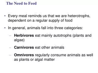
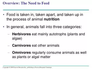
Overview: The Need to Feed • Food is taken in, taken apart, and taken up in the process of animal nutrition • In general, animals fall into three categories: • Herbivores eat mainly autotrophs (plants and algae) • Carnivores eat other animals • Omnivores regularly consume animals as well as plants or algal matter
Nutrition • Nutrition is the process by which organisms use food to perform their life activities. • Nutrients – food chemicals necessary for life. • Metabolism – the breaking down of food for nutrients.
Diet • Provides chemical energy, to be converted into ATP. • Source of organic carbon and organic nitrogen. • Essential nutrients are required by cells and must be obtained from dietary sources
Essential Nutrients • There are four classes of essential nutrients: • Essential amino acids • Essential fatty acids • Vitamins • Minerals
Proteins • Proteins are made up of building blocks called amino acids. • Animals require 20 amino acids and can synthesize about half from molecules in their diet • Essential amino acids, must be obtained from food in preassembled form
Proteins • Protein is very important part of body tissue (especially muscle). • Too little protein causes problems with constructing body tissue. Too much can cause kidney failure. • Rabbit Starvation comes from eating too much lean meat (like rabbit) and not obtaining enough fats and carbohydrates for energy. You will always feel hungry, but carbs and fats are the only things that will satisfy that hunger.
Fig. 41-2 Essential amino acids for adults Beans and otherlegumes Methionine Valine Threonine Phenylalanine Leucine Corn (maize)and other grains Isoleucine Tryptophan Lysine
Essential Fatty Acids • Animals can synthesize most of the fatty acids they need • The essential fatty acids are certain unsaturated fatty acids that must be obtained from the diet • Deficiencies in fatty acids are rare
Vitamins • Vitamins are organic molecules required in the diet in small amounts • 13 vitamins essential to humans have been identified • Vitamins are grouped into two categories: fat-soluble and water-soluble
Minerals • Minerals are simple inorganic nutrients, usually required in small amounts
Dietary Deficiencies • Undernourishment is the result of a diet that consistently supplies less chemical energy than the body requires • Malnourishment is the long-term absence from the diet of one or more essential nutrients • Overnourishment causes obesity, which results from excessive intake of food energy with the excess stored as fat
Ingestion is the act of eating • Digestion is the process of breaking food down into molecules small enough to absorb • Absorption is uptake of nutrients by body cells • Elimination is the passage of undigested material out of the digestive compartment
Fig. 41-7 Smallmolecules Piecesof food Chemical digestion(enzymatic hydrolysis) Nutrientmoleculesenter bodycells Mechanicaldigestion Undigestedmaterial Food Ingestion Digestion Elimination Absorption 2 4 1 3
Fig. 41-9 Crop Gizzard Intestine Esophagus Pharynx Anus Mouth Typhlosole • This digestive tube is called a complete digestive tract or an alimentary canal Lumen of intestine (a) Earthworm Foregut Midgut Hindgut Esophagus Rectum Anus Crop Mouth Gastric cecae (b) Grasshopper Stomach Gizzard Intestine Mouth Esophagus Crop Anus (c) Bird
The mammalian digestive system consists of an alimentary canal and accessory glands. • Mammalian accessory glands are the salivary glands, the pancreas, the liver, and the gallbladder
Food is pushed along by peristalsis, rhythmic contractions of muscles in the wall of the canal • Valves called sphincters regulate the movement of material between compartments
Fig. 41-10a Tongue Sphincter Oral cavity Salivary glands Pharynx Esophagus Sphincter Liver Stomach Ascendingportion oflarge intestine Gall-bladder Duodenum ofsmall intestine Pancreas Smallintestine Smallintestine Largeintestine Rectum Anus Appendix Cecum
Fig. 41-10b Salivaryglands Mouth Esophagus Gall-bladder Stomach Smallintestine Liver Pancreas Largeintestine Rectum Anus A schematic diagram of thehuman digestive system
The Oral Cavity, Pharynx, and Esophagus • Salivary glands deliver saliva to lubricate food • Teeth chew food into smaller particles that are exposed to salivary amylase, initiating breakdown of glucose polymers
The tongue shapes food into a bolus and provides help with swallowing • pharynx, a junction that opens to both the esophagus and the trachea (windpipe) • The esophagus conducts food from the pharynx down to the stomach by peristalsis
Fig. 41-11-3 Food Epiglottisup Tongue Epiglottisup Pharynx Esophagealsphinctercontracted Epiglottisdown Glottis Glottisdownand open Esophagealsphinctercontracted Larynx Trachea Esophagus Esophagealsphincterrelaxed Glottis upand closed Relaxedmuscles Tolungs Tostomach Contractedmuscles Relaxedmuscles Sphincterrelaxed Stomach
Digestion in the Stomach • The stomach stores food and secretes gastric juice, which converts a meal to acid chyme • Gastric juice is made up of hydrochloric acid and the enzyme pepsin • Cells secrete inactive pepsinogen,which is activated to pepsin when mixed with hydrochloric acid in the stomach • Mucus protects the stomach lining from gastric juice
Fig. 41-12 Esophagus Sphincter Stomach Sphincter 5 µm Small intestine Folds ofepithelialtissue Interior surfaceof stomach Epithelium 3 Pepsinogen and HClare secreted. 1 Pepsinogen Pepsin 2 HCl Gastric gland 2 HCl convertspepsinogen to pepsin. 1 3 Pepsin activatesmore pepsinogen. Mucus cells H+ Cl– Chief cells Chief cell Parietal cells Parietal cell
Digestion in the Small Intestine • The small intestine is the longest section of the alimentary canal • It is the major organ of digestion and absorption of water
Fig. 41-13a Carbohydrate digestion Polysaccharides Disaccharides (sucrose, lactose) (starch, glycogen) Oral cavity,pharynx,esophagus Salivary amylase Smaller polysaccharides,maltose Stomach Polysaccharides Lumen ofsmall intestine Pancreatic amylases Maltose and otherdisaccharides Disaccharidases Epitheliumof smallintestine(brushborder) Monosaccharides
Fig. 41-13b Protein digestion Proteins Pepsin Stomach Small polypeptides Polypeptides Pancreatic trypsin andchymotrypsin Lumen ofsmall intestine Smallerpolypeptides Pancreatic carboxypeptidase Amino acids Small peptides Epitheliumof smallintestine(brushborder) Dipeptidases, carboxypeptidase,and aminopeptidase Monosaccharides Amino acids
Fig. 41-13c Nucleic acid digestion DNA, RNA Pancreaticnucleases Lumen ofsmall intestine Nucleotides Nucleotidases Nucleosides Epitheliumof smallintestine(brushborder) Nucleosidasesandphosphatases Nitrogenous bases,sugars, phosphates
Fig. 41-13d Fat digestion Fat globules Bile salts Lumen ofsmall intestine Fat droplets Pancreatic lipase Glycerol, fattyacids, monoglycerides
The first portion of the small intestine is where acid chyme from the stomach mixes with digestive juices from the pancreas, liver, gallbladder, and the small intestine itself
Fig. 41-14 Liver Gallbladder Bile Stomach Secretinand CCK – Gastrin + CCK + Pancreas Duodenum ofsmall intestine Secretin + Key StimulationInhibition CCK + + –
Pancreatic Secretions • The pancreas produces proteases, protein-digesting enzymes that are activated after entering the small intestine • Its solution is alkaline and neutralizes the acidic chyme
Bile Production by the Liver • In the small intestine, bile aids in digestion and absorption of fats • Bile is made in the liver and stored in the gallbladder
Absorption in the Small Intestine • The small intestine has a huge surface area, due to villi and microvilli that are exposed to the intestinal lumen • The enormous microvillar surface greatly increases the rate of nutrient absorption
Fig. 41-15 Microvilli (brushborder) at apical(lumenal) surface Vein carrying bloodto hepatic portal vein Lumen Bloodcapillaries Epithelialcells Basal surface Muscle layers Largecircularfolds Epithelial cells Villi Lacteal Key Lymphvessel Nutrientabsorption Villi Intestinal wall
Absorption in the Large Intestine • The colon of the large intestine is connected to the small intestine
A major function of the colon is to recover water that has entered the alimentary canal • Wastes of the digestive tract, the feces, become more solid as they move through the colon • Feces pass through the rectum and exit via the anus
Energy Sources and Stores • Animals store excess calories primarily as glycogen in the liver and muscles • Energy is secondarily stored as adipose, or fat, cells • When fewer calories are taken in than are expended, fuel is taken from storage and oxidized
Fig. 41-21 Stimulus:Blood glucoselevel risesafter eating. Homeostasis:90 mg glucose/ 100 mL blood Stimulus:Blood glucoselevel dropsbelow set point.
The complexity of weight control in humans is evident from studies of the hormone leptin • Mice that inherit a defect in the gene for leptin become very obese
Fig. 41-24 EXPERIMENT Obese mouse with mutantob gene (left) next to wild-typemouse. RESULTS
Fig. 41-23 Ghrelin Insulin Leptin PYY