Download
1 / 26
280 likes | 787 Views
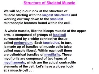
Structure of Skeletal Muscle We will begin our look at the structure of muscle starting with the largest structures and working our way down to the smallest microscopic features found within the cell. A whole muscle, like the biceps muscle of the upper

E N D
Structure of Skeletal Muscle We will begin our look at the structure of muscle starting with the largest structures and working our way down to the smallest microscopic features found within the cell. A whole muscle, like the biceps muscle of the upper arm, is composed of groups of fasciculi surrounded by a white connective tissue called perimysium. Each fasciculus, in turn, is made up of bundles of muscle cells (also called muscle fibers). Within each cell there are cylindrical bundles of myofibrils. These myofibrils are composed of two types of myofilaments, which are the actual contractile elements of the cell. Let's have a closer look at a muscle cell . . .
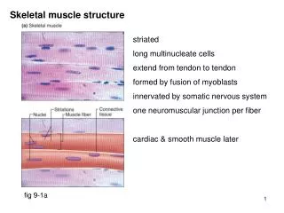
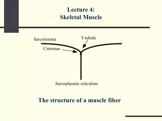
Structure of Skeletal Muscle Muscle cells (or fibers) are one of the few cells in the body with more than one nucleus. They are surrounded by the sarcolemma—the muscle cell membrane—over which the action potential is transmitted. The sarcolemma has small tube-like projections called transverse (T) tubules that extend down into the cell.
Structure of skeletal muscle These T tubules conduct the action potential deep into the cell where the contractile proteins are located. Within the muscle cell are long cylindrical myofibrils that contain the contractile proteins of the muscle.
Structure of skeletal muscle The myofibrils are surrounded by the sarcoplasmic reticulum (SR). This is a mesh-like network of tubes containing Ca++, which are essential for contraction. At either end of and continuous with the SR is the terminal cisterna, which is close to the T tubule where the action potential travels. Let’s now have a look at the contractile proteins found in the myofibrils.
Thin Myofilaments The myofibrils contain two types of myofilaments. The thin myofilaments are composed predominantly of the globular protein actin. Each actin molecule contains a special binding site for the other contractile protein myosin. Many actin molecules are strung together like the beads on a necklace and then twisted to form the backbone of the thin myofilaments.
Thin myofilaments Also found on the thin myofilaments are long protein strands called tropomyosin. When the muscle is at rest, the proteins cover the binding sites for the head of myosin. A third regulatory protein, calledtroponin, is made up of three subunits—troponin , which binds with Ca++; which binds to the tropomyosin; which then exposes the binding site on actin. At rest, the troponin holds the tropomyosin over the myosin binding sites. As we will see later, when Ca++ bind to the troponin unit, the tropomyosin is pulled off the myosin binding sites by the troponin.
Thick Myofilaments The second type of myofilament—the thick myofilament—is made up of the protein myosin. This protein has a long, bendable tail and two heads that can each attach to the myosin binding sites on actin (as mentioned on the previous page). The heads also have a site that can bind and split adenosine triphosphate (ATP). As we will see, it is the splitting of ATP that releases energy to the myosin that powers the contraction of the muscle.
Thick Myofilaments The second type of myofilaments the thick myofilament- is made of the protein myosin. This protein has a long bendable tail and two heads that can each attach to the myosin binding sites on actin. The heads also have a site that can bind and split adenosine triphosphate. As we will see, it is the splitting of ATP that releases energy to the myosin that powers the contraction of the muscle. Many myosin molecules are arranged to form one thick filament. Under a microscope, the arrangement of the thin and thick myofilaments gives the myofibril and the muscle cell a banded appearance. This is why skeletal muscle is called striated muscle.
Actin / Myosin Relationship Groups of thin (actin) myofilaments and groups of thick (myosin) myofilaments are arranged in a repeating pattern (thick, thin, thick, thin, and so on) along the length of the myofibril from one end of the cell to the other. Each group of thin myofilaments extends outward in opposite directions from a central Z line, where they are anchored.
Actin/myosin relationship Similarly, groups of thick myofilaments extend outward from a central M line, where they are attached. Each myofilament is parallel to the length of the myofibril and the muscle cell. The region from one Z line to another is called a sarcomere. This is the smallest functional contractile unit of the muscle cell.
Excitation-contraction Excitation-contraction this is the process by which an action potential in the sarcolemma excites the muscle cell to produce a muscle contraction. The AP that was generated at the neuromuscular junction will spread out over the sarcolemma and down the T-tubules into the core of the muscle. This AP reaches the SR and increases CA++, which will bind to troponin causing tropomyosin to move exposing the binding sites on actin. Myosin will now be able to attach to the actin and a power stroke will occur.
Relaxation of Muscle Once action potentials stop, Ca++ will no longer diffuse out of the sarcoplasmic reticulum (SR). Special calcium pumps rapidly pump Ca++ back into the SR, up its concentration gradient; this requires ATP. Without the calcium present in the cytoplasm of the muscle cell, the tropomyosin will cover the myosin binding sites once again. Myosin will be unable to bind to actin and power strokes will not occur. The muscle will relax.
Actin-Myosin and ATP Cycle Here are the steps summarized 1. Myosin, which has been energized by the splitting of ATP to adenosine diphosphate (ADP) and inorganic phosphate (Pi), attaches to actin* and forms a crossbridge. 2. A power stroke is initiated while ADP and Pi are expelled from the myosin head. 3. Actin and myosin slide past one another.
Actin-Myosin: 4. Actin and myosin are bound together until a new molecule of ATP attaches to myosin; the crossbridge is broken. 5. ATP is split to form ADP and Pi,energizing the myosin molecule. 6. The cycle repeats as long as actin and myosin can interact.
The Motor Unit A motor unit is a motor neuron and all of the muscle cells/fibers it contacts. In almost all situations, one motor neuron will contact (or innervate) several muscle cells,but each muscle cell is innervated by only one motor neuron. The number of muscle cells innervated by a motor neuron varies. A large motor unit has a motor nerve in contact with a large number of muscle cells, while a small motor unit is one in which the motor neuron contacts a few muscle cells.
The Structure of the Neuromuscular Junction The neuron that contacts a muscle cell is sometimes called a motor nerve fiber. The membrane of the axon terminal contains Ca++ voltage-gated channels. These channels open in response to a nerve impulse. The axon terminal of the motor cell/fiber also contains synaptic vesicles that contain the neurotransmitter acetylcholine(ACh).
Neuromuscular -JCT The basement membrane of the axon terminal contains the enzyme acetylcholinesterase (AChE). The muscle cell membrane (also called the sarcolemma) directly under the axon terminal is thrown into folds. This region is called the end plate. The end plate contains receptors for ACh, which are associated with gated ion channels. The gap between the motor fiber and muscle cell is called the synaptic cleft.
Events at the Neuromuscular 1.The action potential on the motor nerve fiber triggers Ca++ voltage-gate channels to open. Ca++ flow into the cell, down their concentration gradient. 2.Ca++ trigger the fusing of synaptic vesiclesto the membrane and the release of ACh into the synaptic cleft by exocytosis. 3.ACh diffuses across the synaptic cleft and attaches to receptors on the muscle cell membrane/sarcolemma. 4.The ACh is broken down by the enzyme AChE and is taken back into the axon terminal to be recycled.