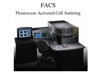
Advanced Applications of FACS in Flow Cytometry for Cell Analysis
260 likes | 369 Views
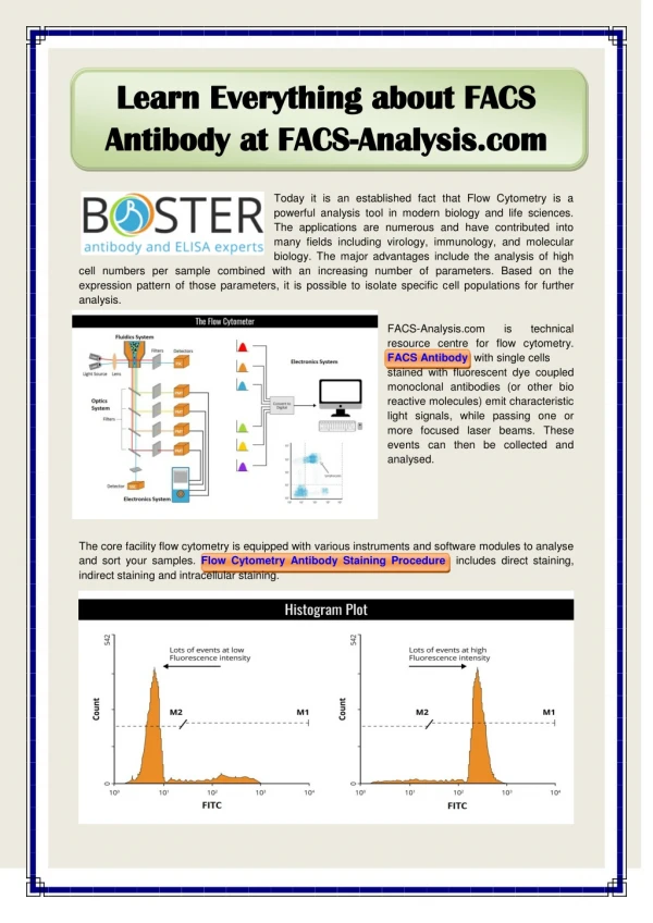
Flow Cytometry using Fluorescence Activated Cell Sorting (FACS) provides powerful methodologies for analyzing and sorting cells based on a variety of measurable parameters. Key applications include evaluating cell volume, morphological complexity, DNA and RNA analysis, protein expression, and cell surface antigens. FACS also enables the assessment of intracellular conditions, apoptosis, cell viability, and drug resistance, particularly in cancer research. This technology remains pivotal for diverse biological studies, including vaccine research and stem cell analysis.

Advanced Applications of FACS in Flow Cytometry for Cell Analysis
E N D
Presentation Transcript
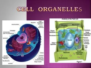
Measurable parameters in flow cytometry • volume and morphological complexity of cells • cell pigments • DNA (cell cycle analysis, cell kinetics, proliferation etc.) • RNA • chromosome analysis and sorting (library construction, chromosome paint) • proteins • cell surface antigens (CD markers) • intracellular antigens (various cytokines, secondary mediators etc.) • nuclear antigens • enzymatic activity • pH, intracellular ionized calcium, magnesium, membrane potential • membrane fluidity • apoptosis (quantification, measurement of DNA degradation, mitochondrial membrane potential, permeability changes) • cell viability • monitoring electropermeabilization of cells • oxidative burst • characterising multi-drug resistance (MDR) in cancer cells • glutathione • various combinations (DNA / surface antigens etc.)
Side scatter http://www.epa.gov/owow/volunteer/proceedings/sixth/session1.html
Side scatter vs Forward scatter University of Utah
fluorimeteroptik Vaccine Research Center / 40 Convent Drive / Bethesda, Maryland 20892
Flourescens colours Fluorescein
Apoptotic cells As cells die or become apoptotic the refractive index of the internal cytoplasm becomes more similar to that of the extracellular medium - this manifests itself as a reduction in forward scatter signal. At the same time, intracellular changes and invagination of the cytoplasmic membrane lead to an increase in side (or orthogonal or 90°) scatter. If we add a dead cell discriminatory dye we can identify cells that have become permeable. In this way we can get low level resolution of dead and apoptotic cells. science.cancerresearchuk.org/.../images/51998
endotelceller R2= total cells R1= living cells Isolation of lymph endothelial cells (LEC) from dermal skin capillary ECs a case study with cells from one donor pre-selected by CD31 magneto bead sorting www.reliatech.de