Connective tissue
Connective tissue. Functions of CT: a. Structural support b. A medium for exchange (so it is vascular). c. Helps in defense & protection of the body. d. A site for storage of fat. TYPES: 1 .CT. proper. 2 . Embryonic CT. 3 . Specialized CT (cartilage, bone and blood).

Connective tissue
E N D
Presentation Transcript
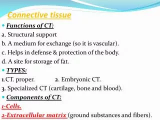
Connective tissue • Functions of CT: a. Structural support b. A medium for exchange (so it is vascular). c. Helps in defense & protection of the body. d. A site for storage of fat. • TYPES: 1.CT. proper. 2. Embryonic CT. 3. Specialized CT (cartilage, bone and blood). • Components of CT: 1-Cells. 2-Extracellular matrix (ground substances and fibers).
Extracellular Matrix I-Ground substance It is a hydrated, amorphous material that is composed of: Glycosaminoglycans, proteoglycans and adhesive glycoproteins as laminin, chondronectin, osteonectin and fibronectin. II-Fibers *Collagen fibers, are inelastic and possess great tensile strength. Each fiber is composed of fine subunits called tropocollagen molecule. • Most of the fibers show axial periodicity by EM. • There are six major collagen types: Type I: in CT proper, bone, dentin and cementum
Type II: In hyaline and elastic cartilage. • Type III: reticular fibers. • Type IV: Lamina densa of the basal lamina.It does not possess a periodicity and not assembled into fibers. • Type V: Associated with type I collagen and in the placenta. • Type VII: Attaching the basal lamina to the lamina reticularis. • The previous types of collagen show fibrillar structure except type IV that form meshwork. *Elastic fibers: Are composed of elastin and microfibrils.They are formed by fibroblasts as well as smooth muscle cells of blood vessels.
CT.Cells • I-Fixed cells: 1.Fibroblasts 2.Adipose cells. 3.Pericytes. 4.Mast cells. 5.Macrophages. • II-Transient cells: 1.Plasma cells. 2.Lymphocytes. 3.neutrophils, eosinophils and basophils. 4.Macrophages. 5.Monocytes.
Fixed CT. Cells I-Fibroblasts They are the most abundant CT. cells and derived from undifferentiated mesenchymal cells. • Types: 1- Active fibroblasts: are elongated, fusiform cells with pale-staining cytoplasm rich in RER and dark-stained large ovoid nucleus containing well-defined nucleolus. 2- Inactive fibroblasts (fibrocytes): are smaller and are more ovoid with more acidophilic cytoplasm. * Fibrobalsts are responsible for the synthesis of almost of extracellular matrix.
II-Adipose cells (fat cells or adipocytes): • They are derived from undifferentiated mesenchymal cells. • They are fully differentiated and do not undergo cell division. • There are 2 types: A-Unilocular fat cells, form white adipose tissue: they are large cells, they store fat as one droplet, which enlarge pushing the cytoplasm and the nucleus peripherally against cell membrane (signet-ring appearance). They have few mitochondria. The fat droplet is not bounded by a membrane. B-Multilocular adipocytes, form brown adipose tissue: Are small cells with multiple fat droplets, central spherical nucleus and many mitochondria. * They function in the synthesis, storage and release of fat.
IV.Mast cells: • They are derived from bone marrow stem cells. • Their cytoplasm is rich in membrane-bound granules that stained metachromatically with toluidine blue. • The granules contain heparin, histamine, eosinophil chemotactic factor and neutrophil chemotactic factor. • They are present in subepithelial CT.,CT. around small blood vessels, and subepithelial CT. of digestive and respiratory tracts (mucosal mast cells that secrete histamine). * Histamine causes vasodilatation and increase permeability of blood vessels. It causes bronchospasm and increase mucus production.
V-Macrophages • They are active mononuclear phagocytes. • Some are fixed and others are transient . • They are irregular in shape and have filopodia. • Their cytoplasm Is basophilic with small eccentric indented nucleus, prominent RER, well developed Golgi and an abundance of lysosomes. • They derived from monocytes that localized in different regions, EX. in liver (Kupffer cells), in lung (dust cells), in CT.(macrophage), in skin (Langerhans cells) and in blood (monocytes). • Several monocytes can fuse together forming foreign-body giant cell. • They phagocytose foreign substances and debris.
Transient CT. cells • I-Plasma cells • They are derived from B-lymphocytes after exposure to an antigen. • They secrete antibodies. • They are large ovoid cells with intensely basophilic cytoplasm that is rich in RER and Golgi (pale-staining region adjacent to the nucleus that has chromatin radiating out from the center (clock-face appearance).
II-Leukocytes • They are white blood cells that circulate in blood stream then migrate through capillaries to enter CT. during inflammations, invasion by foreign elements and immune response.
Classification of CT. A-Embryonic CT: 1.Mesenchymal CT: Is present only in the embryo (in adult in pulp of teeth) and consists of mesenchymal cells in a gel-like ground substance containing scattered reticular fibers. Mesenchymal cells are small cells with pale staining cytoplasm with small processes and a small pale nucleus with prominent nucleolus. 2.Mucous tissue: • It is formed of hyaluronic acid, collagen types I &III and fibroblasts. • It is known as Wharton’s jelly and is found in umblical cord and sub-dermal CT. of the embryo
B-Connective tissue proper 1- Loose (areolar CT): • It fills in the spaces of the body just deep to the skin, below the mesothelium lining of internal body cavity, adventitia of blood vessels and surrounds the parenchyma of glands. • The loose CT of mucous membranes is called lamina propria. • It is characterized by abundant ground substance and tissue fluid housing the fixed CT. cells, undifferentiated cells and collagen, reticular and elastic fibers.
2.Reticular CT: Type III collagen is its major component. Collagen forms mesh-like networks interspersed with fibroblast and macrophages. It forms the architectural framework of liver sinusoids, adipose tissue, lymph nodes, spleen, smooth muscle and islets of Langerhans . 3.Adipose tissue:It is divided into white (unilocular) adipose tissue and brown (multilocular) adipose tissue. It is rich in blood vessels
4. Dense CT: • It has many more fibers and fewer cells than ordinary CT.It is formed of three types: a.Dense irregular collagenous CT: it is formed of randomly arranged collagen fibers, few ground substances and fibroblasts scattering between collagen fibers. It constitutes the dermis of skin, the sheath of nerves and capsules of spleen, testes, ovary and lymph nodes.
b. Dense regullar collagenous CT: it is composed of coarse collagen bundles that oriented into parallel sheets. It has few ground substances and fibroblasts between collagen bundles.Ex. Tendons, ligaments and aponeuroses.
c.Dense regular elastic CT: it has coarse branching elastic fibers with only collagen fibers forming networks and scattered fibroblasts. Elastic fibers are arranged parallel to one another and form either thin sheets or fenestrated membranes as in large blood vessels, ligamenta flava and suspensory ligament of penis.
Basement membrane • It is a cellular region that interface between epithelium and the underlying CT. It is visible by light microscope and stained by PAS. • External lamina, is similar to basement membrane and surrounds smooth muscle, skeletal muscle, adipocytes and Schwann cells. • By EM. It is composed of: 1. Basal lamina which is formed by the epithelium and it is consists of two layers : A. Lamina lucida. It consists of extracellular glycoproteins laminin and entactin. B. Lamina densa, comprises a meshwork of type IV collagen.
2. Lamina reticularis: which is composed of type I &III collagen that manufacture by fibroblasts of underlying CT. It is thick in skin and very thin around alveoli.