Exploring Cortical Activity Changes in Stroke Patients during Robotic Upper Limb Rehabilitation
This study investigates changes in cortical activity among stroke patients, focusing on upper limb rehabilitation through robot-assisted therapy. Via EEG measurements, the protocol examines event-related desynchronization and synchronization during reaching movements. The research aims to clarify neuroplastic changes in the brain post-stroke, utilizing a standardized, natural movement paradigm. Two groups—stroke patients and healthy participants—will undergo similar rehabilitation sessions. Findings aim to enhance understanding of neuroplasticity and inform rehabilitation practices tailored to individual needs.

Exploring Cortical Activity Changes in Stroke Patients during Robotic Upper Limb Rehabilitation
E N D
Presentation Transcript
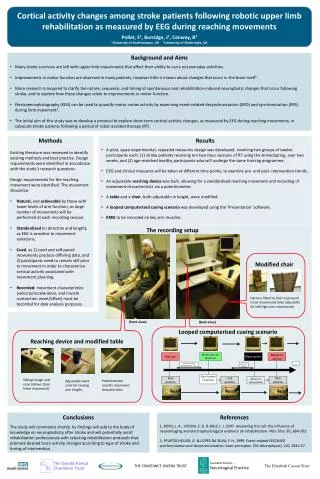
Movement cue: green cue Return to red cue Red cue Black screen … Automatic random interval between 7-11 s 4+ second interval Start forward movement Rest position End position Rest position Return to rest position Cortical activity changes among stroke patients following robotic upper limb rehabilitation as measured by EEG during reaching movements Pollet, S1, Burridge, J1, Conway, B2 1University of Southampton, UK 2University of Strathclyde, UK • Background and Aims • Many stroke survivors are left with upper limb impairments that affect their ability to carry out everyday activities. • Improvements in motor function are observed in many patients, however little is known about changes that occur in the brain itself1. • More research is required to clarify the nature, sequence, and timing of spontaneous and rehabilitation-induced neuroplastic changes that occur following stroke, and to explore how these changes relate to improvements in motor function. • Electroencephalography (EEG) can be used to quantify motor cortex activity by examining event-related desynchronisation (ERD) and synchronisation (ERS) during limb movement2. • The initial aim of this study was to develop a protocol to explore short-term cortical activity changes, as measured by EEG during reaching movements, in subacute stroke patients following a period of robot-assisted therapy (RT). • Methods • Existing literature was reviewed to identify existing methods and best practice. Design requirements were identified in accordance with the study’s research questions. • Design requirements for the reaching movement were identified. The movement should be: • Natural, and achievable by those with lower levels of arm function, as large number of movements will be performed at each recording session. • Standardised (in direction and length), as EEG is sensitive to movement variations. • Cued, as 1) cued and self-paced movements produce differing data, and 2) participants need to remain still prior to movement in order to characterise cortical activity associated with movement planning. • Recorded: movement characteristics (velocity/acceleration, and muscle contraction onset/offset) must be recorded for data analysis purposes. • Results • A pilot, quasi-experimental, repeated measures design was developed, involving two groups of twelve participants each: (1) stroke patients receiving ten two-hour sessions of RT using the ArmeoSpring, over two weeks, and (2) age-matched healthy participants who will undergo the same training programme. • EEG and clinical measures will be taken at different time points, to examine pre- and post- intervention trends. • An adjustable reaching device was built, allowing for a standardised reaching movement and recording of movement characteristics via a potentiometer. • A table and a chair, both adjustable in height, were modified. • A looped computerised cueing scenario was developed using the ‘Presentation’ software. • EMG to be recorded on key arm muscles. The recording setup Modified chair Harness fitted to chair to prevent trunk movements (also adjustable for left/right arm movements) (front view) (back view) Looped computerised cueing scenario Reaching device and modified table Sliding trough and cone (allows 15cm linear movement) Potentiometer records movement characteristics Adjustable hand cone for varying arm lengths Conclusions The study will commence shortly. Its findings will add to the body of knowledge on neuroplasticity after stroke and will potentially assist rehabilitation professionals with selecting rehabilitation protocols that promote desired brain activity changes according to type of stroke and timing of intervention. References 1. BOYD, L. A., VIDONI, E. D. & DALY, J. J. 2007. Answering the call: the influence of neuroimaging and electrophysiological evidence on rehabilitation. PhysTher, 87, 684-703. 2. PFURTSCHELLER, G. & LOPES DA SILVA, F. H. 1999. Event-related EEG/MEG synchronization and desynchronization: basic principles. Clin Neurophysiol, 110, 1842-57. The Gerald Kerkut Charitable Trust Specialist Section – Neurological Practice The Elizabeth Casson Trust THE CONSTANCE OWENS TRUST