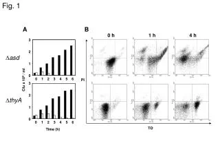
Fig. 45-1
Fig. 45-1. Chapter 45. Hormones and the Endocrine System. Types of Secreted Signaling Molecules. Secreted chemical signals include Hormones : Endocrine signals Local regulators Neurotransmitters Neurohormones Pheromones. Exocrine glands. Fig. 45-2a. Blood vessel. Response.

Fig. 45-1
E N D
Presentation Transcript
Fig. 45-1 Chapter 45 Hormones and theEndocrine System
Types of Secreted Signaling Molecules • Secreted chemical signals include • Hormones :Endocrine signals Local regulators • Neurotransmitters • Neurohormones • Pheromones Exocrine glands
Fig. 45-2a Blood vessel Response (a) Endocrine signaling Response (b)Paracrine signaling act on cells near the secreting cell Response (c) Autocrine signaling:signals act on the secreting cell itself
Fig. 45-2b Synapse Neuron Response (d) Synaptic signaling Neurosecretory cell Blood vessel Response (e) Neuroendocrine signaling
Fig. 45-3 Water-soluble Lipid-soluble Chemical Classes of Hormones 0.8 nm Polypeptide: Insulin Steroid: Cortisol Amine: Epinephrine Amine: Thyroxine
Hormone Receptor Location: Scientific Inquiry • In the 1960s, researchers studied the accumulation of radioactive steroid hormones in rat tissue • These hormones accumulated only in target cells that were responsive to the hormones • These experiments led to the hypothesis that receptors for the steroid hormones are located inside the target cells • Further studies have confirmed that receptors for lipid-soluble hormones such as steroids are located inside cells
Fig. 45-4 1.The hormone triggered a response only when it was allowed to bind to cell surface receptors 2. This confirmed that water-soluble receptors were on the cell surface RESULTS MSH injected into melanocyte Melanocyte with melanosomes (black dots) Melanosomes do not disperse Melanosomes disperse Nucleus MSH injected into interstitial fluid (blue)
Fig. 45-5-1 Cellular Response Pathways Fat-soluble hormone Water- soluble hormone Transport protein Signal receptor TARGET CELL Signal receptor NUCLEUS (a) (b)
Fig. 45-5-2 Fat-soluble hormone Water- soluble hormone Transport protein Signal receptor TARGET CELL OR Signal receptor Cytoplasmic response Gene regulation Cytoplasmic response Gene regulation NUCLEUS (a) (b)
Pathway for Water-Soluble Hormones Fig. 45-6-1 Epinephrine Adenylyl cyclase G protein GTP G protein-coupled receptor ATP Second messenger cAMP
Fig. 45-6-2 Epinephrine Adenylyl cyclase G protein GTP G protein-coupled receptor ATP Second messenger cAMP Protein kinase A Inhibition of glycogen synthesis Promotion of glycogen breakdown
Fig. 45-7-1 Hormone (estradiol) Pathway for Lipid-Soluble Hormones Estradiol (estrogen) receptor Plasma membrane Hormone-receptor complex
Fig. 45-7-2 Hormone (estradiol) Estradiol (estrogen) receptor Plasma membrane Hormone-receptor complex DNA Vitellogenin mRNA for vitellogenin
VitellogeninFrom Wikipedia, the free encyclopediaJump to: navigation, search Vitellogenin (VTG or less popularly known as VG) (from latin vitellus = yolk and gener = to produce) is a synonymous term for the gene and the expressed protein. The protein molecule is classified as a glyco-lipo-protein, having properties of a sugar, fat and protein. Vitellogenin is an egg yolk precursor protein expressed in females of fish, amphibians, reptiles (including birds), insects and the platypus. In the presence of estrogenic endocrine disruptive chemicals (EDCs), male fish can express the Vg gene in a dose dependent manner. Vg gene expression in male fish can be used as a molecular marker of exposure to estrogenic EDCs.
Multiple Effects of Hormones • The same hormone may have different effects on target cells that have • Different receptors for the hormone • Different signal transduction pathways • Different proteins for carrying out the response • A hormone can also have different effects in different species
Fig. 45-8-1 Same receptors but different intracellular proteins (not shown) Epinephrine Epinephrine receptor receptor Glycogen deposits Vessel dilates. Glycogen breaks down and glucose is released. (a) Liver cell (b) Skeletal muscle blood vessel
Fig. 45-8-2 Same receptors but different intracellular proteins (not shown) Different receptors Epinephrine Epinephrine Epinephrine Epinephrine receptor receptor receptor receptor Glycogen deposits Vessel dilates. Vessel constricts. Glycogen breaks down and glucose is released. (a) Liver cell (c) Intestinal blood vessel (b) Skeletal muscle blood vessel
Signaling by Local Regulators • Types of local regulators: • Cytokines and growth factors • Nitric oxide (NO) • Prostaglandins: • Prostaglandins help regulate aggregation of platelets, an early step in formation of blood clots
Concept 45.2 • Negative feedback and antagonistic hormone pairs are common features of the endocrine system
Fig. 45-10 Major endocrine glands: Hypothalamus Pineal gland Pituitary gland Organs containing endocrine cells: Thyroid gland Thymus Parathyroid glands Heart Liver Adrenal glands Stomach Pancreas Kidney Testes Small intestine Kidney Ovaries
Fig. 45-11 Pathway Example – Stimulus Low pH in duodenum S cells of duodenum secrete secretin ( ) Endocrine cell Negative feedback Blood vessel Target cells Pancreas Bicarbonate release Response
Fig. 45-12-5 Body cells take up more glucose. Insulin and Glucagon: Insulin Beta cells of pancreas release insulin into the blood. Liver takes up glucose and stores it as glycogen. STIMULUS: Blood glucose level rises. Blood glucose level declines. Homeostasis: Blood glucose level (about 90 mg/100 mL) STIMULUS: Blood glucose level falls. Blood glucose level rises. Alpha cells of pancreas release glucagon. Liverbreaks downglycogen andreleases glucose. Glucagon
Diabetes Mellitus • Diabetes mellitus is perhaps the best-known endocrine disorder • Type I diabetes mellitus (insulin-dependent) is an autoimmune disorder in which the immune system destroys pancreatic beta cells • Type II diabetes mellitus (non-insulin-dependent) involves insulin deficiency or reduced response of target cells due to change in insulin receptors
Concept 45.3 :Coordination of Endocrine and Nervous Systems in Invertebrates • In insects, molting and development are controlled by a combination of hormones: • A brain hormone stimulates release of ecdysone from the prothoracic glands • Juvenile hormone promotes retention of larval characteristics • Ecdysone promotes molting (in the presence of juvenile hormone) and development (in the absence of juvenile hormone) of adult characteristics
Fig. 45-13-3 Brain Neurosecretory cells Corpus cardiacum PTTH Corpus allatum Low JH Prothoracic gland Ecdysone Juvenile hormone (JH) EARLYLARVA LATER LARVA PUPA ADULT
Coordination of Endocrine and Nervous Systems in Vertebrates • The hypothalamus receives information from the nervous system and initiates responses through the endocrine system • Attached to the hypothalamus is the pituitary gland composed of the posterior pituitary and anterior pituitary
Fig. 45-14 Cerebrum Thalamus Pineal gland Hypothalamus Cerebellum Pituitary gland Spinal cord Hypothalamus Posterior pituitary Anterior pituitary
Fig. 45-15 Hypothalamus Posterior Pituitary Hormones Neurosecretorycells of thehypothalamus Axon Posterior pituitary Anterior pituitary HORMONE Oxytocin ADH TARGET Kidney tubules Mammary glands,uterine muscles
Oxytocin induces uterine contractions and the release of milk • Suckling sends a message to the hypothalamus via the nervous system to release oxytocin, which further stimulates the milk glands • This is an example of positive feedback, where the stimulus leads to an even greater response • Antidiuretic hormone (ADH) enhances water reabsorption in the kidneys
Fig. 45-16 Pathway Example Stimulus Suckling + Sensoryneuron Hypothalamus/posterior pituitary Positive feedback Neurosecretorycell Posterior pituitarysecretes oxytocin ( ) Bloodvessel Smooth muscle inbreasts Targetcells Response Milk release
Anterior Pituitary Hormones • Hormone production in the anterior pituitary is controlled by releasing and inhibiting hormones from the hypothalamus • For example, the production of thyrotropin releasing hormone (TRH) in the hypothalamus stimulates secretion of the thyroid stimulating hormone (TSH) from the anterior pituitary
Fig. 45-17 Tropic effects only:FSHLHTSHACTH Neurosecretory cellsof the hypothalamus Nontropic effects only:ProlactinMSH Nontropic and tropic effects:GH Hypothalamicreleasing andinhibitinghormones Portal vessels Endocrine cells ofthe anterior pituitary Posterior pituitary Pituitary hormones HORMONE FSH and LH TSH ACTH Prolactin MSH GH TARGET Testes orovaries Thyroid Adrenalcortex Mammaryglands Melanocytes Liver, bones,other tissues
Hormone Cascade Pathways • A hormone can stimulate the release of a series of other hormones, the last of which activates a nonendocrine target cell; this is called a hormone cascade pathway • The release of thyroid hormone results from a hormone cascade pathway involving the hypothalamus, anterior pituitary, and thyroid gland
Fig. 45-18-3 Pathway Example Hormone cascade pathways are usually regulated by negative feedback Stimulus Cold Sensoryneuron – Hypothalamus secretesthyrotropin-releasinghormone (TRH ) Neurosecretorycell Bloodvessel – Anterior pituitary secretes thyroid-stimulatinghormone (TSHor thyrotropin ) Negative feedback Thyroid gland secretes thyroid hormone (T3 and T4 ) Targetcells Body tissues Increased cellularmetabolism Response
Tropic Hormones • A tropic hormone regulates the function of endocrine cells or glands • The four strictly tropic hormones are • Thyroid-stimulating hormone (TSH) • Follicle-stimulating hormone (FSH) • Luteinizing hormone (LH) • Adrenocorticotropic hormone (ACTH)
Nontropic Hormones • Nontropic hormones target nonendocrine tissues • Nontropic hormones produced by the anterior pituitary are • Prolactin (PRL) • Melanocyte-stimulating hormone (MSH) • Prolactin stimulates lactation in mammals but has diverse effects in different vertebrates • MSH influences skin pigmentation in some vertebrates and fat metabolism in mammals
Growth Hormone • Growth hormone (GH) is secreted by the anterior pituitary gland and has tropic and nontropic actions • It promotes growth directly and has diverse metabolic effects • It stimulates production of growth factors • An excess of GH can cause gigantism, while a lack of GH can cause dwarfism
Concept 45.4 • Endocrine glands respond to diverse stimuli in regulating metabolism, homeostasis, development, and behavior
Thyroid Hormone: Control of Metabolism and Development • The thyroid gland consists of two lobes on the ventral surface of the trachea • It produces two iodine-containing hormones: triiodothyronine (T3) and thyroxine (T4) • Thyroid hormones stimulate metabolism and influence development and maturation • Hyperthyroidism, excessive secretion of thyroid hormones, causes high body temperature, weight loss, irritability, and high blood pressure • Graves’ disease is a form of hyperthyroidism in humans • Hypothyroidism, low secretion of thyroid hormones, causes weight gain, lethargy, and intolerance to cold
Fig. 45-19 High leveliodineuptake Normaliodineuptake
Parathyroid Hormone and Vitamin D: Control of Blood Calcium • Two antagonistic hormones regulate the homeostasis of calcium (Ca2+) in the blood of mammals • Parathyroid hormone (PTH) is released by the parathyroid glands • Calcitonin is released by the thyroid gland
Fig. 45-20-2 Activevitamin D Stimulates Ca2+uptake in kidneys Increases Ca2+ uptake in intestines PTH Parathyroid gland(behind thyroid) Stimulates Ca2+ release from bones STIMULUS: Falling bloodCa2+ level Blood Ca2+level rises. Homeostasis: Blood Ca2+ level(about 10 mg/100 mL)
PTH increases the level of blood Ca2+ • It releases Ca2+ from bone and stimulates reabsorption of Ca2+ in the kidneys • It also has an indirect effect, stimulating the kidneys to activate vitamin D, which promotes intestinal uptake of Ca2+ from food • Calcitonin decreases the level of blood Ca2+ • It stimulates Ca2+ deposition in bones and secretion by kidneys
Adrenal Hormones: Response to Stress • The adrenal glands are adjacent to the kidneys • Each adrenal gland actually consists of two glands: • the adrenal medulla (inner portion) and adrenal cortex (outer portion)
Catecholamines from the Adrenal Medulla • The adrenal medulla secretes epinephrine (adrenaline) and norepinephrine (noradrenaline) • These hormones are members of a class of compounds called catecholamines • They are secreted in response to stress-activated impulses from the nervous system • They mediate various fight-or-flight responses
Catecholamines • They are called catecholamines because they contain a catechol group, and are derived from the amino acidtyrosine.[3] • The most abundant catecholamines are epinephrine (adrenaline), norepinephrine (noradrenaline) and dopamine, all of which are produced from phenylalanine and tyrosine. • Catecholamines are water-soluble and are 50% bound to plasma proteins, so they circulate in the bloodstream. • Tyrosine is created from phenylalanine by hydroxylation by the enzyme phenylalanine hydroxylase. (Tyrosine is also ingested directly from dietary protein). It is then sent to catecholamine-secreting neurons. Here, many kinds of reactions convert it to dopamine, to norepinephrine, and eventually to epinephrine