Light
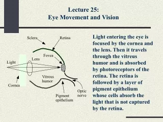
Sclera. Retina. Fovea. Lens. Light. Vitreus humor. Cornea. Optic nerve. Pigment epithelium. Lecture 25: Eye Movement and Vision.

Light
E N D
Presentation Transcript
Sclera Retina Fovea Lens Light Vitreus humor Cornea Optic nerve Pigment epithelium Lecture 25: Eye Movement and Vision Light entering the eye is focused by the cornea and the lens. Then it travels through the vitreus humor and is absorbed by photoreceptors of the retina. The retina is followed by a layer of pigment epithelium whose cells absorb the light that is not captured by the retina.
Low light, poor resolution Outer segment Inner segment Nucleus Synaptic terminal Rod Cone Light Receptors The loss of cones is considered legal blindness. Rods and cones consist of the outer segment (which contains the light-transducing apparatus), the inner segment (which holds the nucleus and much of the biochemical machinery), and the synaptic terminal (which makes connections with the receptor’s target cells).
Rod Cone I Info II Horizontal cell Bipolar cells III Amacrine cell IV V Ganglion cell Optic nerve Light Neurons of the Retina Below the photoreceptor level of the retina, there is an intermediate layer containing three types of cells: bipolar cells, amacrine cells, and horizontal cells. Ganglionic cells are located under the intermediate layer. Their axons form the optic nerve. • I: outer nuclear layer • II: outer plexiform layer • III: inner nuclear layer • IV: inner plexiform layer • V: ganglion cell layer
Point of gaze fixation Left visual hemifield Right visual hemifield Nose Temporal hemiretina Temporal hemiretina Nasal hemiretinas Visual Fields Definitions of the left and right visual hemifields. Note that the optic tracts convey information from the ipsilateral temporal hemiretina and the contralateral nasal hemiretina—i.e., visual information about the contralateral visual hemifield.
Gaze Adduction/abduction Elevation/depression Intorsion/extorsion Eye Rotations
Eye Movements • Saccades: quick jumps; V up to 900°/s • Smooth pursuit: following a target; V < 100°/s • Vergence: changing the depth of fixation
Saccades • Velocity cannot be controlled voluntarily • Feedforward control • Generated in the pontine and midbrain reticular centers, with participation of the cerebral cortex and the basal ganglia
Smooth Pursuit • Requires a moving target • Involved structure: • Striate cortex • Prestriate motor areas • Pons • Cerebellum
Reflex Eye Movements Vestibulo-ocular reflex (VOR): • Brainstem level • Latency of 14 ms • Nystagmus Optokinetic reflex: • Stabilizes image on the retina during head movements • Involves cortical structures • Longer latency
Optic nerve Thalamus (lateral geniculate nucleus) Midbrain (pretectal area) Superior colliculus Visual perception Saccades Pupillary reflexes Optic Nerve Projections The optic nerve projects onto three subcortical areas: the lateral geniculate nucleus of the thalamus, the pretectal area of the midbrain, and the superior colliculus. Projections to the lateral geniculate nucleus participate in visual perception, projections to the superior colliculus control saccades, and projections to the pretectal area control pupillary reflexes.
Ciliary ganglion Edinger-Westphal nucleus Optic nerve Pretectal area The Pupillary Reflex Neurons in the pretectal area receive an input from the optic nerve and project to the Edinger-Westphal nucleus. It, in turn, generates a parasympathetic input to oculomotor neurons in the ciliary ganglion. These neurons innervate the smooth muscle of the pupillary sphincter.
Sensory inputs Maps Visual Superior colliculi Auditory Somatosensory Motor Brain stem Head and neck movements Cerebellum Eye movements Maps in the Superior Colliculi The superior colliculi integrate sensory information from different sources and contain three sensory maps and one motor map. The superior colliculi project to the regions of the brain stem that control eye movements and contribute to two descending tracts: the tectospinal tract (which is involved in the reflex control of head and neck movements) and the tectopontine tract (which delivers visual information to the cerebellum for further processing).
Horizontal connections Input Columns in the Visual Cortex The primary visual cortex is organized into vertical narrow columns running from the surface to the white matter. Each column is approximately 30 to 100 µm wide and 2 mm deep. There is an orderly shift in the axis of orientation from one column to its neighbors. There are horizontal connections among columns so that the activity of a neuron within a column may be influenced by stimuli of other orientations.