fMRI guided Microarray analysis
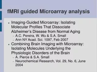
fMRI guided Microarray analysis. Imaging-Guided Microarray: Isolating Molecular Profiles That Dissociate Alzheimer’s Disease from Normal Aging A.C. Pereira, W. Wu & S.A. Small Ann NY Acad. Sci. 1097, Feb 2007

fMRI guided Microarray analysis
E N D
Presentation Transcript
fMRI guided Microarray analysis • Imaging-Guided Microarray: Isolating Molecular Profiles That Dissociate Alzheimer’s Disease from Normal Aging • A.C. Pereira, W. Wu & S.A. Small • Ann NY Acad. Sci. 1097, Feb 2007 • Combining Brain Imaging with Microarray: Isolating Molecules Underlying the Physiologic Disorders of the Brain • A. Pierce & S.A. Small • Neurochemical Research, Vol. 29, No. 6, June 2004
Crash course: The CELL and microarrays in 3 slides • Cells internal processes and inter-cell communication based on proteins • Goal: Figure out which proteins exist in a cell under some condition • Condition – e.g. disease • Many times – detect proteins differentially expressed – e.g. disease vs. control • Basic: staining a specific protein and follow it under a microscope • Next: The CELL
From DNA to Protein • (Final) product – Protein • Intermediate product mRNA • Idea: measure mRNA to get protein measurements • Simultaneous measurements by hybridization
DNA Microarrays • mRNA – concatenation of nucleotides • 4 types ATGC – pegs/holes • Process • Crush cell • Wash all but mRNA • Glue lamps • Spill on chip • Shake well!
Sorry, 4 slides... • Chip design – probes for genes • Light on --> Protein exists • Light off --> No protein at the moment
Problem setting • Given two sets of DNA microarrays: • Disease • Control • Extract a set of differentially expressed genes • Feature selection for classification • Biological significant features for downstream research
Problem setting revisited • Given two sets of DNA microarrays: • Disease • Control • + fMRI measurements of the two populations • Extract a small set of differentially expressed pathogenic-behaving genes • Feature selection for classification • Biological significant features for downstream research
Nervous System Diseases • Multiple categorizations: • Organic vs. Functional • Anatomic vs. Physiologic • Structural vs. Metabolic • Physiologic = molecular pathway • Invisible to (non functional) imaging • Not evident under microscope, no histological markers • Anatomic = loss/gain of tissue
A Needle in a Haystack • Target: Find the one(?) molecule that malfunctions: • Multiple molecular pathways within a neuron • Neuronal interconnection • Cascade/ripple throughout the system • Molecule -> Neuron (population) • Neuron -> Other neuron • Other neuron -> Other molecules • Molecules might be in the same neuron population (feedback) • infeasible for standard statistical analysis
Aging and AD • Cognitive decrease (AD and aging) • Differential – vulnerable vs. resistant • Memory Encoding • Hippocampus • Entorhinal Cortex • Dentate Gyrus • CA subfields • Subiculum • Common process: • Synaptic Failure leads to: • Cell loss / tangles / plaques • Function, not structure!
Hippocampus • AD • Aging • Known from postmortem,in-vitro, and fMRI • Interconn. • Asses all regions together
Microarray analysis • Differential expression analysis • “Blind” analysis • Thousands of parameters simultaneously • High false positives rate (multiple comparisons, recall FDR) • Poor signal-to-noise ratio • Usually produce a “list” of differentially expressed genes • “list” can be very long (up to hundreds)
Statistical Modeling • Temporal model • 2nd stage for fMRI • Double subtraction • With sickness - basal metabolic rate changes as well
Multiple Studies • Why fMRI and not postmortem? • p.m. biased against earliest (and most discriminatory) stages • Only fMRI can image the cell-sickness stage • EC found to be the primary source of dysfunction in AD • What about normal aging? • Age-related changes in the EC matched pathological decline • Age-related changes in the dentate gyrus (DG), and subiculum (SUB), matched normal aging
Spatio-Temporal Model • How a pathogenic molecule should behave? • Differentially expressed in the EC (vs. no differentially expression in the DG) • Differences between AD and controls should be age independent • once EC dysfunction begins it does not worsen across age groups or over time
Results • 5 Molecules matched the pattern • Much less than 100s! • Best molecule: VPS35 • Part of a complex that connects-to and transports substances within a cell • A-beta – a known “smoking gun” for AD • Experiments validated: • Low VPS35 --> High A-beta • Required neuronal molecules in end-to-end transportation are not transported --> brain dysfunction
Conclusion • Microarrays – noisy, unfocused results • fMRI – imaging in-vivo, not post-mortem • Create statistical model (criteria) using fMRI, for microarray differentiation • Lack of specific methods • Not a parametric model, like a thumb rule • Nice example for research advance • My personal research is on PD • Lots of imaging data • Any suggestions? • Thanks!