Download
1 / 64
640 likes | 759 Views
Structure databases, searches and alignments. Marian Novotny marian@xray.bmc.uu.se. Molecular Bioinformatics X3. Outline. Structure databases - why do we need them? - types of structural databases

E N D
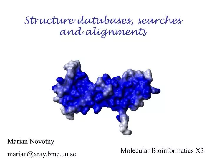
Structure databases, searches and alignments Marian Novotny marian@xray.bmc.uu.se Molecular Bioinformatics X3
Outline Structure databases - why do we need them? - types of structural databases - Protein Data Bank - other useful databases Searches - text searches Structure searches (alignments) - why? - how ? - comparison of available tools
Structure databases Why? • data tend to get lost • source of information for further analysis • better access to data by general public • validation of data is (sometimes) possible
Database is… …. a structured collection of data held in computer storage; esp. one that incorporates software to make it accessible in a variety of ways; transf., any large collection of information. Oxford English dictionary …..a usually large collection of data organized especially for rapid search and retrieval (as by a computer) Merriam-Webster Online
Databases Primary databases Derived databases Added-value databases RCSB MSD PDBJ NDB CSD OCA PDBSum EDS Whatcheck Jena Image library
Primary databases • repositories of experimental data of macromolecular structures (X-ray, NMR, electron microscopy…) • RCSB (USA), MSD (Europe) and PDBJ (Japan) collaborate to form wwPDB. Data can be submitted to any of these databases. Databases interchange their new data on a regular basis, so they have an identical content. • primary databases differ in presentation of data and the amount of extra services and links they provide
The Protein Data Bank (PDB) • established in 1971 by Walter Hamilton at Brookhaven National Laboratory • seven structures were deposited at the beginning • the database was distributed on magnetic tapes • RCSB now run by the consortium of three institutions (San Diego Supercomputer Centre, Rutgers University and Centre for Avanced Reasearch and Biotechnology) • 29326 structures (26.01.2005) • distributed over internet • released once a week
HEADER HYDROLASE 27-OCT-03 1UR9 TITLE INTERACTIONS OF A FAMILY 18 CHITINASE WITH THE DESIGNED TITLE 2 INHIBITOR HM508, AND ITS DEGRADATION PRODUCT, TITLE 3 CHITOBIONO-DELTA-LACTONE COMPND MOL_ID: 1; COMPND 2 MOLECULE: CHITINASE B; COMPND 3 CHAIN: A, B; COMPND 4 EC: 3.2.1.14; COMPND 5 ENGINEERED: YES; COMPND 6 MUTATION: YES SOURCE MOL_ID: 1; SOURCE 2 ORGANISM_SCIENTIFIC: SERRATIA MARCESCENS; SOURCE 3 STRAIN: BJL200; SOURCE 4 EXPRESSION_SYSTEM: ESCHERICHIA COLI; SOURCE 5 EXPRESSION_SYSTEM_STRAIN: DH5 ALPHA; SOURCE 6 OTHER_DETAILS: CLONED GENE KEYWDS CHITINASE, INHIBITION, LACTONE, CHITIN DEGRADATION, KEYWDS 2 HYDROLASE, GLYCOSIDASE EXPDTA X-RAY DIFFRACTION AUTHOR G.VAAJE-KOLSTAD,A.VASELLA,M.G.PETER,C.NETTER,D.R.HOUSTON, AUTHOR 2 B.WESTERENG,B.SYNSTAD,V.G.H.EIJSINK,D.M.F.VAN AALTEN REVDAT 1 27-APR-04 1UR9 0 JRNL AUTH G.VAAJE-KOLSTAD,A.VASELLA,M.G.PETER,C.NETTER, JRNL AUTH 2 D.R.HOUSTON,B.WESTERENG,B.SYNSTAD,V.G.H.EIJSINK JRNL AUTH 2 D.M.F.VAN AALTEN JRNL TITL INTERACTIONS OF A FAMILY 18 CHITINASE WITH THE JRNL TITL 2 DESIGNED INHIBITOR HM508 AND ITS DEGRADATION JRNL TITL 3 PRODUCT, CHITOBIONO-DELTA-LACTONE JRNL REF J.BIOL.CHEM. V. 279 3612 2004 JRNL REFN ASTM JBCHA3 US ISSN 0021-9258 REMARK 1 REMARK 1 REFERENCE 1 REMARK 1 AUTH D.M.F.VAN AALTEN,D.KOMANDER,B.SYNSTAD,S.GASEIDNES, REMARK 1 AUTH 2 M.G.PETER,V.G.H.EIJSINK REMARK 1 TITL STRUCTURAL INSIGHTS INTO THE CATALYTIC MECHANSIM OF REMARK 1 TITL 2 A FAMILY 18 EXOCHITINASE REMARK 1 REF PROC.NAT.ACAD.SCI.USA V. 98 8979 2001 REMARK 1 REFN ASTM PNASA6 US ISSN 0027-8424 REMARK 1 REFERENCE 2 REMARK 1 AUTH D.M.F.VAN AALTEN,B.SYNSTAD,M.B.BRURBERG,E.HOUGH, REMARK 1 AUTH 2 B.RIISE,V.G.H.EIJSINK,R.K.WIERENGA REMARK 1 TITL STRUCTURE OF A TWO-DOMAIN CHITOTRIOSIDASE FROM PDB FILE
PDB file format Chain Atom type Occupancy Atom identifier 12345678901234567890123456789012345678901234567890123456789012345678901234567890 1 2 3 4 5 6 7 8 ATOM 340 N PHE A 43 3.853 28.346 32.161 1.00 10.57 N ATOM 341 CA PHE A 43 3.839 29.688 32.724 1.00 12.33 C ATOM 342 C PHE A 43 3.096 29.747 34.047 1.00 13.20 C ATOM 343 O PHE A 43 2.361 28.823 34.393 1.00 12.52 O ATOM 344 CB PHE A 43 3.228 30.659 31.700 1.00 10.99 C ATOM 345 CG PHE A 43 3.993 30.709 30.401 1.00 9.80 C ATOM 346 CD1 PHE A 43 3.743 29.794 29.386 1.00 9.85 C ATOM 347 CD2 PHE A 43 5.032 31.615 30.233 1.00 11.37 C ATOM 348 CE1 PHE A 43 4.528 29.781 28.220 1.00 10.71 C ATOM 349 CE2 PHE A 43 5.816 31.612 29.075 1.00 10.61 C ATOM 350 CZ PHE A 43 5.569 30.697 28.067 1.00 10.48 C Atom number Residue number X,Y,Z coordinates Temperature factor Residue type
1234567890123456789012345678901234567890123456789012345678901234567890123456789012345678901234567890123456789012345678901234567890123456789012345678901234567890 1 2 3 4 5 6 7 8 ATOM 340 N PHE A 43 3.853 28.346 32.161 1.00 10.57 N ATOM 341 CA PHE A 43 3.839 29.688 32.724 1.00 12.33 C ATOM 342 C PHE A 43 3.096 29.747 34.047 1.00 13.20 C ATOM 343 O PHE A 43 2.361 28.823 34.393 1.00 12.52 O ATOM 344 CB PHE A 43 3.228 30.659 31.700 1.00 10.99 C ATOM 345 CG PHE A 43 3.993 30.709 30.401 1.00 9.80 C ATOM 346 CD1 PHE A 43 3.743 29.794 29.386 1.00 9.85 C ATOM 347 CD2 PHE A 43 5.032 31.615 30.233 1.00 11.37 C ATOM 348 CE1 PHE A 43 4.528 29.781 28.220 1.00 10.71 C ATOM 349 CE2 PHE A 43 5.816 31.612 29.075 1.00 10.61 C ATOM 350 CZ PHE A 43 5.569 30.697 28.067 1.00 10.48 C ATOM 340 N PHE A 43 3.853 28.346 32.161 1.00 10.57 N ATOM 341 CA PHE A 43 3.839 29.688 32.724 1.00 12.33 C ATOM 342 C PHE A 43 3.096 25.747 34.047 1.00 13.20 C ATOM 343 O PHE A 43 2.361 28.823 34.393 1.00 12.52 O ATOM 344 CB PHE A 43 3.228 30.659 31.700 1.00 10.99 C ATOM 345 CG PHE A 43 3.993 30.709 30.401 1.00 9.80 C ATOM 346 CD1 PHE A 43 4.743 29.794 29.386 1.00 9.85 C ATOM 347 CD2 PHE A 43 5.032 31.615 30.233 1.00 11.37 C ATOM 348 CE1 PHE A 43 4.528 32.781 28.220 1.00 10.71 C ATOM 349 CE2 PHE A 43 5.816 31.612 29.075 1.00 10.61 C ATOM 350 CZ PHE A 43 5.569 30.697 28.067 1.00 10.48 C
PDB files - problems • PDB format uses fixed-width fields, so one entry is limited to 99,999 atom records and chain identifier is limited to single character (not even for structures of huge complexes - e.g. ribosome and viruses) • 12345678901234567890123456789012345678901234567890123456789012345678901234567890 • 1 2 3 4 5 6 7 8 • ATOM 340 N PHE A 43 3.853 28.346 32.161 1.00 10.57 N - parsing of PDB files difficult - apart from ATOM records the file is almost unstructured (e.g. no rules to describe structure determination in REMARKS records) mmCIF and XML formats deal with these issues
Trust PDB? The database centres can’t refuse to accept any data! Even if curators of the PDB know the data contain serious errors. So, PDB does contain a lot of errors - from sequence consistency errors (you’ll deal with them) to completely wrong folds. And even the best data are still only the models that fit best experimental data. Never trust the PDB!
Do you find this Trp normal? Trp D 67 7GPB
Validation of structure files • - check statistics for bond lengths, angles, Ramachandran plots…. • - do statistics look similar to those of other proteins? • WhatCheck, Procheck • how well does the model fit experimental data? • EDS
Text searches in structural databases Find all the structures deposited by Gerard Kleywegt with resolution better than 2Å and published in Journal of Molecular Biology Options: PDB - SearchLite, SearchFields MSD - MSDlight, MSDpro (Java), MSDmine OCA
Summary • - three major repositories of structural data: RCSB, MSD and PDBJ • all three are part of wwPDB • structural data are deposited in PDB files - problems • new formats - mmCIF, XML • validation tools are necessary - WHATCheck, EDS • new services are developed to analyze the whole database (MSD services) • searches at various levels of depth/complexity - Searchlite, Search Fields • added-value databases - OCA, PDBSum
Why structural alignment ? we have sequence alignment - Clustal… KTHLCV KSHA-V that gives us an idea about a correspondence of amino acids of two (or more ) proteins That enables to infer information about function And evolution of the Protein If the sequences are similar enough !!!!
What is twilight zone ? Sequence alignment unambiguously distinguishes only between protein pairs of similar structure and non-similar structures when the pairwise sequence identity is high. High sequence identity roughly means over 40 %. The signal gets blurred in the twilight zone of 20-35 % sequence identity.
More of the twilight zone More than 90 % sequence pairs with the sequence identity lower than 25 % have different structures. Significance of sequence alignments is length dependent. The longer the sequence the lower identity is required to be called significant.Nevertheless, it converges to 25% with alignments longer than 80 amino acids. ‘The more similar than identical’ rule can reduce a number of false positives. Using intermediate sequences for finding links between more distant families can also reduce the number of false positives.
How far can the sequence identity drop? Average sequence identity of random alignments - 5.6 % Average sequence identity of remote homologues 8.5 %
How does it work? From http://www.biochem.unizh.ch/antibody/Introduction/Institutsseminar97/source/slide2.htm
Structural alignment because: Structures are better conserved than sequences structural alignment can imply a functional similarity that is not detectable from a sequence alignment . Might help to improve sequence alignment when structures are available (phylogenetic studies, homology modeling). Will improve sequence alignment methods (use of structural alignments’ substitution matrices, gap penalties). Will improve sequence prediction methods
Structural versus sequence alignment 1FWR_A -------------------------MKNWKTSAESILTTGPVVPVIVVKKLEHAVPMAKA 2YPI_A ARTFFVGGNFKLNGSKQSIKEIVERLNTASIPENVEVVICPPATYLDYSVSLVKKPQVTV ::. . . : :. * .. : . * ... 1FWR_A LVAGGVRVLEVTLRTECAVDAIRAIAKEVPEAIVGAGTVLNPQQLAEVTE-------AGA 2YPI_A GAQNAYLKASGAFTGENSVDQIKDVGAKWVILGHSERRSYFHEDDKFIADKTKFALGQGV . .. . :: * :** *: :. : . :: ::: *. 1FWR_A QFAISPGLTEPLLKAATEGTIPLIPGISTVSELMLGMDYGLKEFQFFPAEANGGVKALQA 2YPI_A GVILCIGETLEEKKAGKTLDVVERQLNAVLEEVKDWTNVVVAYEPVWAIGTGLAATPEDA . :. * * **.. : :.:.*: : : .:. :. .... :* 1FWR_A IAGPFSQVRFCPKGGISPANYRDYLALKSVLCIGGSWLVPADALEAGDYDRITKLAREAV 2YPI_A QDIHASIRKFLASKLGDKAASELRILYGGSANGSNAVTFKDKADVDGFLVGGASLKPEFV * :* .. . * . : . ..: . .* * :.* * * 1FWR_A EGAKL-- 2YPI_A DIINSRN Sequence 1 ------------ART---FFVGGNFKLNG-SKQSI-KEIVERLNTASI--PENVEVVICP .=ALI |=ID | |.... .. ..... . ....|... . | ... Sequence 2 MKNWKTSAESIL--TTGP--VVPVI--VVKKLEHAVP-MAKALVAG-GVR-----V-LEV Sequence 1 ------PATYLDYSVSLV-KKPQVTVGAQ-N--AY-LKASGAFTGEN-S---VDQIKDVG .=ALI |=ID ...........| . ..|||. . . . . . .| Sequence 2 TLRTECAVDAIRAIAKEVP-E--AIVGAGTVLN-PQ----------QLAEVT--E---AG Sequence 1 AKWVILGH--SERRSYFHEDDKFIADKTKFALGQGVGVILCIGETLEEKKAGKTLDVVER .=ALI |=ID |...|. . .....|.|.......|..|. ... Sequence 2 AQFAIS-PGL-------------TEPLLKAATEGTIPLIPGIS--------------TVS Sequence 1 QLNAV-LEEVKDW-TNVVVAYEP--VW--AIGTGLAATPEDA--QDI--HASI-RKFLA- .=ALI |=ID .|... . .. . .....| . . . . .. . . Sequence 2 ELMLGMD--YG-LK---EFQFFPAE-ANG-------G----VKA--LQA--IAG-P--FS Sequence 1 SKLGDKAA-SELRILYGGSANGSN-AVTF---KDK-ADVDGFLVGGA-SLK--------- .=ALI |=ID . |....|... .. . . . ..|..... .. .. Sequence 2 -------QV---RFCPKGGIS-PANY--RDYL--ALKSVLCIGG-SWL-VPADALEAGDY Sequence 1 --P--EFV--DIIN--SR-N .=ALI |=ID . . . . . .. Sequence 2 DRITKL-AREA--VEGAKL-
Sequence versus structural alignment 2 3 4 5 6 7 8 9 10 11 12 13 14 PHE ASP ILE CYS ARG LEU PRO GLY SER ALA GLU ALA VAL CYS PHE ASN VAL CYS ARG THR PRO --- --- --- GLU ALA ILE CYS PHE ASN VAL CYS ARG --- --- --- THR PRO GLU ALA ILE CYS
Is it difficult to make structural alignment? Structural alignment is NP-hard (nondeterministic polynomial time) problem. In other words, it is not tractable properly. Even, if it would, the result would be correct from technical point of view not necessary from biological point of view. Yes, it is.
General solution Use a heuristic approach: Represent the proteins A and B in some coordinate independent space Compare A and B Optimize the alignment between A and B (e.g. minimize R.M.S.d.) Measure the statistical significance of the alignment against some random set of structure comparisons
“..in some coordinate independent space…” • Make the problem easier by: • comparing only distance matrices of atoms • comparing secondary • structure element (SSE) • comparing cartoons • comparing vectors of SSE • combination of mentioned methods • ….
None of the methods guarantee the finding of the closest structure and two methods can disagree at all amino acid positions. Nevertheless they can still provide a valuable insight into the history of the protein and give hints concerning the function.