Download
1 / 105
1.09k likes | 1.69k Views
Myasthenia Gravis. Disease of the neuromuscular junction characterized by fluctuating weakness of certain skeletal muscle groups. Myasthenia Gravis(MG). Acetycholine (ACh) is an important neurotransmitter that stimulates muscle tissue to contract.

E N D
Myasthenia Gravis • Disease of the neuromuscular junction characterized by fluctuating weakness of certain skeletal muscle groups.
Myasthenia Gravis(MG) • Acetycholine (ACh) is an important neurotransmitter that stimulates muscle tissue to contract. • MG is an autoimmune disease in which antibodies are formed against ACh and a reduction in ACh receptor sites at the neuromuscular junction.
Pathophysiology • Loss of muscle strength. • There is no single cause identified, however, thymic tumors and viral infections have been found in a certain number of patients.
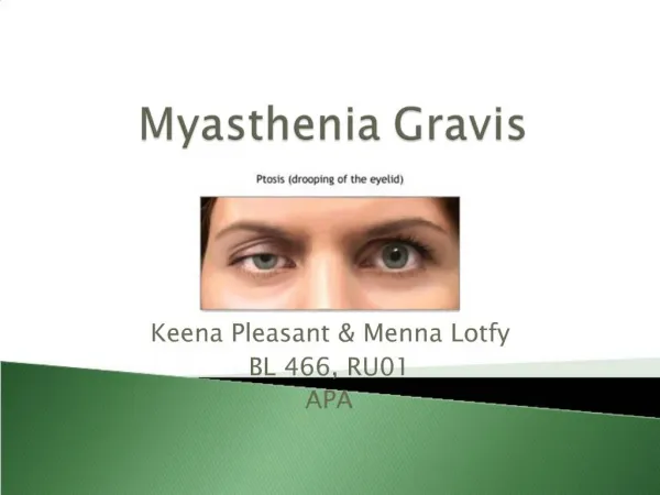
Clinical manifestations • Primary s/s= easy fatigability of skeletal muscle during activity. • Muscles involved: eyes and eyelids, chewing, swallowing, speaking, and breathing. • Fluctuating weakness: usually strong in the a.m., progressively weaker with activity.
Clinical Manifestations • 90% of patients have eye involvement • Facial mobility may be impaired • Muscles of limb and trunk less often affected. • No sensory or reflex loss; muscle atrophy is rare.
Clinical manifestations • Variable course • May be precipitated by emotional stress, pregnancy, menses, secondary illness, trauma, temperature extremes, hypokalemia, ingestion of drugs with neuromuscular blocking agents, surgery.
Complications • Aspiration, respiratory insufficiency, and respiratory infection • Acute exacerbation called myasthenic crisis. • The opposite of this is a cholinergic crisis and results from overdose of cholinergic drugs.
Diagnostic studies • Assessment: • Have pt look up for 2-3 minutes; if MG, patient will have increased droop of eyelids. • EMG may show muscle fatigue • Tensilon test- in MG reveal improved muscle contractility after IV anticholinesterase agent edrophonium chloride (tensilon) • Also diagnosis cholinergic crisis- muscle weakness gets worse • Keep atropine on hand to counteract effects of tensilon
Therapeutic management • Anticholinesterase inhibitors-prevents anticholinestersase from breaking down ACh; helps neurotransmission. Monitor dose! • Mestinon, Prostigmine Corticosteroids- decrease immune response Prednisone Plasmapheresis- removes ACh antibodies and short-term improvement.
Nursing Management • See handout!
ALS • Rare progressive disease characterized by loss of motor neurons. • Onset between 40 and 70 years of age • Twice as many men as women
ALS • Motor neurons degenerate and leave dead tissue which cannot send signals from brain to muscles • Weakness of upper extremities, dysarthria, dysphagia • Muscle wasting • Death from respiratory infection DT compromised respiratory function; usually w/in 2-6 years.
ALS • No cure • No definitive test; diagnosis made through physical exam and series of tests which rule out other diseases which look like ALS. • EMG and nerve conduction studies may be done. • Patient still cognitively intact!
Drug Therapy • Riluzole (Rilutek)- delays progression of ALS. • Decreases amount of glutamate (an excitatory neurotransmitter) in the brain. • May delay need for trach and death by a few months.
Nursing care • Support cognitive and emotional functions • Facilitate communication • Provide diversional activities (read) • Help family and pt with advanced planning (hospice) • Provide companionship
Trigeminal neuralgia • Also called tic douloureax • Uncommon cranial nerve disorder • More common in women @ 50-60 years of age • Trigeminal nerve is 5th cranial nerve (CNV) • And has both motor and sensory branches; mostly maxillary and mandibular branches involved.
Trigeminal Neuralgia • Initiating pathologic events include: • nerve compression by tortuous arteries of the posterior fossa blood vessels • demyelinating plaques • herpes virus infection • infection of teeth and jaw • a brainstem infarct
Clinical manifestations • Abrupt onset with excruciating pain!! • Pain described as burning, knifelike, or lightinglike shock in the lips, upper or lower gums, cheek, forehead, or side of the nose. • Patient may twitch, grimace, frequent blinking and tearing of eye (tic) may occur.
Clinical manifestations • Attacks may be brief (2 or 3 minutes) • Unilateral • Episodes may be initiated by triggering mechanism of light cutaneous stimulation as a specific point (trigger zone) along nerve branches.
Precipitating stimuli • Chewing, brushing teeth, hot or cold blast of air on the face, washing the face, yawning, or talking. • Patient may eat improperly, neglect hygiene practices, wear cloth over face, withdraw from interaction with others.
Diagnostic studies • Need to rule out other neurological causes of facial and cephalic pain. • CT scan will rule out brain lesions, vascular malformations. LP and MRI will r/o MS. • There is no specific diagnostic test for TN.
Drug Therapy • Antiseizure meds may prevent and acute attack or promote remission-mechanism unknown. • Carbamazeprine (Tegretol)…most common • Phenytoin (Dilantin) • Valproate (Depakene)
Drug Therapy • Carbamazeprine has side effects: • Bone marrow suppression leading to blood abnormalities (CBC counts needed) • Pain relief not permanent
Conservative Therapy • Nerve block • Biofeedback
Surgery • Percutaneous radiofrequency rhizotomy (electrocoagulation)- placing a needle into the trigeminal nerve to destroy the area by radiofrequency currents. • May lose corneal reflex • Easily performed; minimal risk • Pain relieved, but face is numb • (Table 57-2, Lewis, 1715)
Microvascular decompression • Most common surgical procedure • Blood vessels that are compressing the nerve are displaced and repositioned. This relieves pain without residual sensory loss. • Long-term success rate • Safe without residual sequelae • Recurrence occurs in 30% of patients within 6 years
Glycerol rhizotomy • Injecting glycerol near the root of the nerve • Safer and fewer risks that percutaneous types.
Nursing Interventions • See handout • Pay special attention to nursing implementation section in Lewis (1716-1717).
Bell’s Palsy • Characterized by: • Peripheral facial paralysis • Acute benign cranial polyneuritis Acute disorder characterized by a disruption of the motor branches of cranial nerve VII on one side of the face. (in absence of stroke)
Can affect any age group, though more common from 20-60. Etiology unknown; though reactivated herpes simplex may be involved. Reactivation causes edema, inflammation, ischemia, and eventual demyelination of the nerve, creating pain and alteration in motor and sensory function. Bell’s Palsy
Benign, with 85% of people recovering in 6 months-remaining 15% have some asymmetry of facial muscles Clinical manifestations
Clinical manifestations • Often accompanied by an outbreak of herpes vesicles in or around the ear. • Pain around or behind the ear • Fever, tinnitus, hearing deficits • Flaccidity of the affected side of the face with drooping of the mouth accompanied by drooling DT paralysis of the facial nerve (motor branches)
Clinical manifestations • Inability to close the eyelids, with an upward movement of the eyeball when closure is attempted; lower lid may turn out • Wide palpebral fissure (opening between eyelids) • Flattening of the nasolabial fold • Inability to smile, frown, or whistle • Unilateral loss of taste • Altered chewing ability; loss of or excessive tearing
Complications • Psychological withdrawal DT changes in appearance,malnutrition or dehydration, mucous membrane trauma, corneal abrasion, muscle stretching, and facial spasms and contractures.
Diagnostic Studies • Diagnosis made on basis of symptoms in the absence of other causes of paralysis such as stroke. • No definitive test • EMG may determine nerve excitability or absence
Therapeutic Management • Corticosteroids- drug of choice • Prednisone may be started immediately! • Best if initiated before paralysis is complete • Taper off over 2 weeks • Decrease edema and pain Analgesics may be needed for pain Antivirals : Acyclovir (Zovirax) and Famvir because HSV is implicated in 70% of cases. See Lewis 1719-1720- Nursing Implementation
Guillain-Barre’ Syndrome • Post-infectious polyneuropathy; ascending polyneuropathic paralysis • An acute, rapidly progressing and potentially fatal form of polyneuritis
Guillain-Barre’ Syndrome • Affects the peripheral nervous system
Guillain-Barre’ • T-cell sensitization occurs which causes loss of myelin which disrupts nerve impulses • Loss of myelin, edema and inflammation of the affected nerves, causes a loss of neurotransmission to the periphery. • 85% of patients recover with supportive care.
Pathophysiology • Etiology unknown • May be cell-mediated immunological reaction directed at the peripheral nerves • Frequently preceded by viral infection, trauma, surgery or other immune system stimulation.
Clinical Manifestations • Usually develop 1 to 3 weeks after URI or GI infection • Weakness of lower extremities (symmetrically) • Parathesia (numbness and tingling), followed by paralysis • Hypotonia and areflexia (absence of reflexes) • Pain in the form of muscles cramps or hyperesthesias (worse at night).