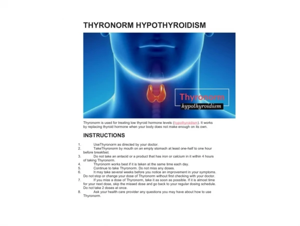

Hypothyroidism
E N D
Presentation Transcript
Hypothyroidism DR LALEH GHANEI ASSISTANT PROFESSOR OF ENDOCRINOLOGY
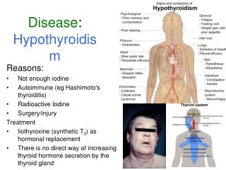
Chronic autoimmune thyroiditis-goitrous & atrophic forms Radiation Surgery Infiltrative disease(amyloidosis,sclerodermia) Iodine deficiency Biosynthetic defect Drugs (lithium,iodine,contrast agents) Pituitary disease Hypothalamic disease Causes of hypothyroidism Primary Secondary
Chronic Autoimmune Thyroiditis Hashimoto's thyroiditis (chronic autoimmune thyroiditis) is the most common cause of hypothyroidism in iodine-sufficient areas of the world. gradual thyroid failure, goiter, or both, due to autoimmune-mediated destruction of the thyroid gland. Nearly all patients have high serum concentrations of antibodies against one or more thyroid antigens, lymphocytic infiltration of the thyroid, which includes thyroid-specific B and T cells, and apoptosis of thyroid follicular cells.
Incidence of hypothyroidism* Female 40/10,000 Male 6/10,000 Prevalence of Hypothyroidism* Female %9.3 Male %1.3 *2779 people in UK with a medium age of 58 years Whickham survey 1995
Spectrum of thyroid autoimmunity Grave’s Disease Hashimoto Disease Postpartum thyroiditis Silent thyroiditis Drug induced thyroiditis
Possible precipitating factors • Genetic • Infection • Stress • Humoral factors (sex steroids , pregnancy)
Pathogenesis • Thyroid auotantigenes • Role of B cells • The primary role of T cells • Potential mechanism of thyroid injury Molecular mimicry Bystander activation Thyroid cell expression of HLA Ag Thyroid cell apoptosis
PATHOLOGY The characteristic histopathological abnormalities are profuse lymphocyticinfiltration, lymphoid germinal centers, and destruction of thyroid follicular cells . fibrosis and areas of follicular-cell hyperplasia, presumably induced by TSH, are also seen in patients with severe disease. The intrathyroidal lymphocytes are both T and B lymphocytes.
Effects of Thyroid Hormone • Fetal brain and skeletal maturation • Increase in basal metabolic rate • Inotropic and chronotropic effects on heart • Increases sensitivity to catecholamines • Stimulates gut motility • Increase bone turnover • Increase in serum glucose, decrease in serum cholesterol
Table 330-5. Signs and Symptoms of Hypothyroidism (Descending Order of Frequency) Symptoms Tiredness, weakness Dry skin Feeling cold Hair loss Difficulty concentrating and poor memory Constipation Weight gain with poor appetite Dyspnea Hoarse voice Menorrhagia (later oligomenorrhea or amenorrhea) Paresthesias Impaired hearing Signs Dry coarse skin; cool peripheral extremities Puffy face, hands and feet (myxedema) Diffuse alopecia Bradycardia Peripheral edema Delayed tendon reflex relaxation Carpal tunnel syndrome Serous cavity effusions
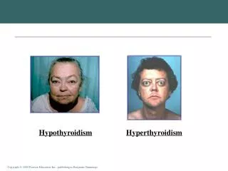
Figure 9-3. (A) The classic torpid facies of severe myxedema in a man. The face appears puffy, and the eyelids are edematous. The skin is thickened and dry. (B) The facies in pituitary myxedema is often characterized by skin of normal thickness, covered by fine wrinkles. Puffiness is usually less than in primary myxedema. The eyelids are often edematous. The palpebral fissure may be narrwowed because of blepharoptosis, due to diminished tone of the sympathetic nervous fibers to Müller's levator palpebral superious muscle and is the opposite of the lid retraction seen in thyrotoxicosis. The modest measurable exophthalmos seen in some patients with myxedema is presumably related to accumulation of the same mucous edema in the orbit as is seen elsewhere. It is not progressive and carries no threat to vision, as in the ophthalmopathy of Graves' disease. The tongue is usually large, occasionally to the point of clumsiness. Sometimes a patient will complain of this problem. Sometimes it is smooth, as in pernicious anemia (of course, pernicious anemia may coexist). Patients do not usually complain of soreness of the tongue, as they may in pernicious anemia. When anemia is marked, the tongue may be pale, but more often it is red, in contrast to the pallid face
TSH and Free T4 TSH Free T4 TSH Free T4 TSH Free T4 TSH Free T4 Central hypo. NTI Drug effect Normal Primary hypothyroidism Mild or Subclinical hypothyroidism
SCREANING • goiter • women older than 60 years (AACE&ATA consensus) • history of autoimmune disease(EX DMI) • previous radioactive iodine therapy • History of head and neck irradiation • family history of thyroid disease • use of medications that may impair thyroid function
SCREANING • symptoms of hypothyroidism • laboratory or radiologic abnormalities that could be caused by hypothyroidism • patients taking drugs that may impair thyroid function
SCREANING • We also suggest universal screening for thyroid dysfunction in pregnant women or those hoping to become pregnant
As examples, thyroid function should be measured in patients with the following: • Substantial hyperlipidemia • Hyponatremia, • High serum muscle enzyme concentrations. • Macrocytic anemia • Pericardial or pleural effusions • Pituitary or hypothalamic disorders • History of autoimmune diseases
three settings in which measurement of serum TSH may not be a useful tool for the diagnosis of hypothyroidism • pituitary or hypothalamic disease is known or suspected. • In hospitalized patients • In patients receiving drugs or with underlying diseases which affect TSH secretion . Drugs that can decrease TSH secretion include dopamine, high doses of glucocorticoids,phenytoin, and somatostatin analogues (such as octreotide). Drugs that increase TSH secretion include dopamine antagonists (metoclopramide or domperidone), amiodarone, and oral cholecystographic dyes (sodium ipodate).
Hypothroidism in adulscent • Hypothyroidism is the most common disturbance of thyroid function in children, and is most often caused by chronic autoimmune thyroiditis • Euthyroidgoiter is more common than hypothyroidism • Hypothyroidism in children can have deleterious effects on growth, pubertal development and school performance.
Hypothroidism in adulscent • The most common physical finding at presentation is a goiter. • The most common manifestation of hypothyroidism in children is declining growth velocity, often resulting in short stature.
Hypothroidism in adulscent • Pubertal development is delayed in most , some children have sexual precocity. • A few patients have hyperprolactinemia and rarely galactorrhea. Growth hormone normal or decreased, and serum insulin-like growth factor I (IGF-I) levels are usually decreased
Hypothroidism in adulscent primary hypothyroidism must be excluded in any child with an enlarged sella turcica. it rarely causes symptoms or signs, unlike a pituitary tumor or craniopharyngioma, and is reversible with T4 therapy.
Hypothroidism in adulscent • Atrophic thyroiditisis primarily the result of cell-mediated cytotoxicity leading to follicular cell apoptosis; • complement-dependent antibody-mediated cytotoxicity may contribute to thyroid damage. • TSH receptor blocking antibodies result in loss of thyroid morphological integrity, which may be reversible.
Hypothroidism in adulscent • Goitrous thyroiditis may be induced by one of three mechanisms: lymphocytic and plasma cell infiltration (and lymphoid germinal centers), • the production of antibodies that stimulate thyroid growth, • or excess TSH secretion.
Hypothroidism in adulscent • Children at increased risk for chronic autoimmune thyroiditis and, to a lesser extent, hypothyroidism. : • Down syndrome (trisomy 21), • Turner syndrome, • type 1 (autoimmune) diabetes mellitus, ( 20 percent have high serum antithyroid antibody concentrations, and 5 percent have abnormalities in thyroid function, usually subclinical hypothyroidism) • celiac disease, and • possibly Klinefelter syndrome
Hypothroidism in adulscent • children with Down, Turner, or Klinefelter syndrome, type 1 diabetes, or celiac disease should be screened annually for hypothyroidism by measuring serum TSH.
DIAGNOSIS • TSH levels have diurnal variation, and one series suggests that TSH values measured at 8 AM are more sensitive for the diagnosis of mild primary hypothyroidism as compared with measurements at 4 PM • Mild elevations of serum TSH (up to 7.5 mU/L), TSH levels are normal in up to 70 percent of patients when the test is repeated . • Thus,Treatment decisions in such patients should be made only after repeat testing.
Pharmacokinetics of oral thyroid hormones T4 T3 Absorption ~ 80% ~90% Time to peak level 2-4 h 1-2h Increase in serum concentration 20-40% 250-600% Half-life 6-7days ~1days
TREATMENT • In most patients, hypothyroidism is a permanent condition requiring lifelong treatment. • Therapy consists of thyroid hormone replacement unless the hypothyroidism is transient (as after painless thyroiditis or subacute thyroiditis) or reversible (due to a drug that can be discontinued).
TREATMENT • The treatment of choice for correction of hypothyroidism is synthetic thyroxine (T4). • Approximately 80 percent of a dose of T4 is absorbed and, because the plasma half-life of T4 is long (seven days), once-daily treatment results in nearly constant serum T4 and triiodothyronine (T3) concentrations when a steady state is reached
TREATMENT • Because there may be subtle differences in bioavailability between T4 formulations, we feel that it is preferable to stay with one formulation when possible. • If the preparation must be changed, follow-up biochemical monitoring should be done to determine if retitration of the dose is necessary. We typically measure a serum TSH six weeks after changing preparations
TREATMENT • Adverse effects of T4 replacement are rare as long as the correct dose is given. • Rare patients have an allergy to the dye or filler in the tablets. For dye sensitivities, multiples of the white 50 mcg tablets can be given.
Dose and monitoring • The average replacement dose of T4 in adults is approximately 1.6 mcg/kg body weight per day (112 mcg/day in a 70-kg adult) • T4 requirements correlate better with lean body mass than total body weightT4 requirements correlate better with lean body mass than total body weight
Average Replacement Dose of T4 • I.6 to 1.8µ/kg ideal body wight/day • 75-125μg/day in women and 125-200μg/day in men • Older patients(>60years) require 20% to 30% less T4 per kg ideal body weight than do younger patients
Timing of dose • T4 should be taken on an empty stomach, ideally an hour before breakfast • T4 should not be taken with other medications that interfere with its absorption, such as bile acid resins, proton pump inhibitors, calcium carbonate, and ferrous sulfate. • ( should be taken several hours after the T4 dose. ) • after initiation of T4 therapy, the patient should be reevaluated and serum TSH measured in six weeks. TSH above the normal reference range, the dose of T4 can be increased by 12 to 25 mcg/day.
Goals of therapy • amelioration of symptoms • normalization of TSH secretion • reduction in the size of the goiter • keep TSH within the normal reference range (approximately 0.5 to 5.0 mU/L). • hypothyroid symptoms, and the TSH at the upper limit, increase the dose and to aim for a serum TSH value in the lower half of the normal range.
It is prudent to keep TSH values in the lower normal range(0.4-2.0 mU/l), and to avoid TSH levels of <0.1 mU/l
Hypothroidism in adulscent • There is some controversy about the need to treat children with mild subclinical hypothyroidism, characterized by TSH elevations between 6 and 10 mU/L. • There is general agreement to treat children with subclinical hypothyroidism and TSH levels >10 mU/L.
Hypothroidism in adulscent • central hypothyroidism in children is characterized by a low ratio of the TSH values taken at 8 AM and 4 PM (8AM:4PM ratio <1.3 in central hypothyroidism; normal 8AM:4PM ratio >1.5), when serum free T4 is in the lower third of the normal range.
Treatment in children • The goals of treatment are to restore normal growth and development, including pubertal development. • Children clear T4 more rapidly than adults; as a result, the daily replacement dose on a weight basis is higher: • Age 1 to 3 years — 4 to 6 mcg/kg body weight • Age 3 to 10 years — 3 to 5 mcg/kg • Age 10 to 16 years — 2 to 4 mcg/kg
Alternatively, the replacement dose can be calculated as a function of body surface area, in which case the dose at any age is approximately 100 mcg/m2/day. Body surface area can be determined from height and weight using a calculator • BSA=sqr(height*weight/3600)
In children with primary hypothyroidism, the recommended target range for TSH is in the lower half of the reference range (optimally 0.5 to 2.0 mU/L) and for T4 or free T4 is in the upper half of the reference range • The optimal T4 range is between 9 and 13 ug/dL (116 to 167 nmol/L) • if the normal free T4 reference range is 0.6 to 1.8 ng/dL (8 to 23 pmol/L), the corresponding optimal free T4 range would be between 1.2 and 1.8 ng/dL (15 to 23 pmol/L).
For most children, therapy should be initiated with a dose in the middle of the appropriate range for age. • However, in those with long-standing hypothyroidism, a somewhat lower dose should be given to avoid overly rapid acceleration of skeletal maturation and loss of adult height. • In one study of children presenting prior to puberty with severe hypothyroidism, the height deficit after treatment ranged from 8 to 14 cm at the end of puberty
Patients with longstanding hypothyroidism are at risk for developing pseudotumor cerebri shortly after initiation of levothyroxine treatment . • Children with persistent headaches or vision changes should contact their physician.