Download
1 / 3
30 likes | 61 Views
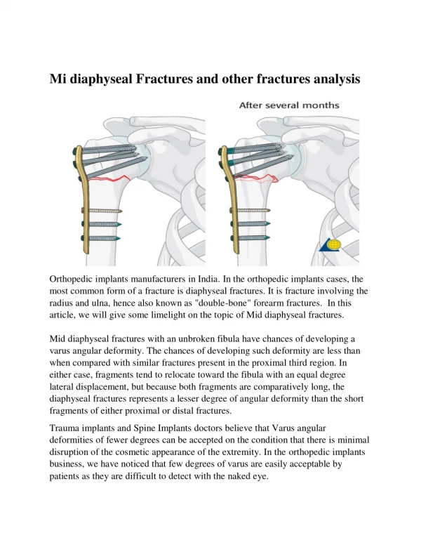
Orthopedic implants manufacturers in India. In the orthopedic implants cases, the most common form of a fracture is diaphyseal fractures. It is a fracture involving the radius and ulna, hence also known as "double-bone" forearm fractures. In this article, we will give some limelight on the topic of Mid diaphyseal fractures.<br><br>Read more: https://www.siiora.lk/locking-plates/

E N D
Mi diaphyseal Fractures and other fractures analysis Orthopedic implants manufacturers in India. In the orthopedic implants cases, the most common form of a fracture is diaphyseal fractures. It is fracture involving the radius and ulna, hence also known as "double-bone" forearm fractures. In this article, we will give some limelight on the topic of Mid diaphyseal fractures. Mid diaphyseal fractures with an unbroken fibula have chances of developing a varus angular deformity. The chances of developing such deformity are less than when compared with similar fractures present in the proximal third region. In either case, fragments tend to relocate toward the fibula with an equal degree lateral displacement, but because both fragments are comparatively long, the diaphyseal fractures represents a lesser degree of angular deformity than the short fragments of either proximal or distal fractures. Trauma implants and Spine Implants doctors believe that Varus angular deformities of fewer degrees can be accepted on the condition that there is minimal disruption of the cosmetic appearance of the extremity. In the orthopedic implants business, we have noticed that few degrees of varus are easily acceptable by patients as they are difficult to detect with the naked eye.
If one considers valgus plane cases, then similar deformities in the valgus plane are more easily recognizable. It is also not possible to correct a varus malformation that develops as a result of an intact fibula by manipulation. However, its degree of severity can most likely be reduced to some extent by proper molding of the initial and subsequent stabilizing devices or the choice of the contour of the orthopedic instruments like a prefabricated brace. The impact can also be reduced by improving the internal rotation of the proximal fragment using an external rotation force to the distal fragment. Orthopedic implant company research and development team also found that the patients who have significant amounts of bowing and internal rotation of the tibia prior to the injury are more probable to develop greater deformities compared to those patients with a straighter tibia. In any case, the start of weight-bearing ambulation (capability to walk without the need for any kind of assistance) for patients with an intact fibula should begin afterward than patients with other fractures. It is done in the hope that the built-in soft tissue stability might lessen the sternness of angular deformity. Orthopedic implants India study found out that varus angulation develops in most fractures of the tibia with an intact fibula and also that these patients must be followed more closely and have X-rays of their extremities taken on a more frequent basis for a detailed analysis. The study further concludes that if the irregularity does not develop beyond 5 marks, its fixedness in the same state can be accepted, as in such case cosmetic appearance and functional performance of the extremity will not be negotiated. However, any further succession of the abnormality calls for the treatment of fibular osteotomy (cutting of bone to relocate alignment) or any other means to correct the alignment like an intramedullary fixation.
Surgeons’ expert on orthopedic surgical instrumentssupplier performs the osteotomy at a level almost reverse to the fractured tibia. Once it is done, the extremity once again can be soothed using an above-the-knee walking cast (for 1-2 weeks) and further transferring it to a functional brace. Our Official Contact Details: Website: https://www.siiora.lk/ Email Id: online@siora.net PH: 9810021264 Name: Anuj Dureja