Cell Culture
l ifetechnologies.com. Cell Culture. What is cell culture?. Removal of cells from an animal or plant and their subsequent growth in a favorable artificial environment Much more difficult than growing established bacterial and fungal strains. Biosafety Levels. Biosafety Level 1 (BSL-1)


Cell Culture
E N D
Presentation Transcript
lifetechnologies.com Cell Culture
What is cell culture? • Removal of cells from an animal or plant and their subsequent growth in a favorable artificial environment • Much more difficult than growing established bacterial and fungal strains
Biosafety Levels • Biosafety Level 1 (BSL-1) • BSL-1 is the basic level of protection common to most research and clinical laboratories, and is appropriate for agents that are not known to cause disease in normal, healthy humans. • Biosafety Level 2 (BSL-2) • BSL-2 is appropriate for moderate-risk agents known to cause human disease of varying severity by ingestion or through percutaneous or mucous membrane exposure. Most cell culture labs should be at least BSL-2, but the exact requirements depend upon the cell line used and the type of work conducted. • Biosafety Level 3 (BSL-3) • BSL-3 is appropriate for indigenous or exotic agents with a known potential for aerosol transmission, and for agents that may cause serious and potentially lethal infections. • Biosafety Level 4 (BSL-4) • BSL-4 is appropriate for exotic agents that pose a high individual risk of life-threatening disease by infectious aerosols and for which no treatment is available. These agents arerestrictedto high containment laboratories.
Equipment Needed • Cell culture hood (i.e., laminar-flow hood or biosafety cabinet) – Need at least Class II • Incubator (humid CO2 incubator recommended) • Water bath • Centrifuge • Refrigerator and freezer (–20°C) • Cell counter (e.g., CountessRII Automated Cell Counter or hemacytometer) • Inverted microscope • Liquid nitrogen (N2) freezer or cryostorage container • Sterilizer (i.e., autoclave) • Aspiration pump (peristaltic or vacuum) • pH meter • Cell culture vessels (e.g., flasks, Petri dishes, roller bottles, multiwell plates) • Pipettes and pipettors • Syringes and needles • Small waste containers • Media, sera, and reagents • Cells
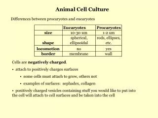
Culture Conditions • Can vary depending on cell type but include: • a substrate or medium that supplies the essential nutrients (amino acids, carbohydrates, vitamins, minerals) • growth factors • Fetal bovine serum is often used – high embryonic growth factors • hormones • gases (O2, CO2) • a regulated physico-chemical environment (pH, osmotic pressure, temperature)
Primary Cell Culture • Cells are isolated from the tissue and start to proliferate • Confluence – occupy all the available substrate (aka bottom of well or flask) • When confluence is reached, cells need to subcultured
After subculture, the primary culture becomes known as a cell line • Finite vs. Continuous Cell Line • Normal cells only divide a certain number of times • If cells go through transformation, they can become immortal or continuous • Can be induced spontaneously, by chemical, or by virus
Media • Type depends on cell type • Contains vitamins, nutrients, antibiotics (in some cases) • Fetal bovine serum used for growth factors • Remember, cells of multicellular organisms rely on other cell types within the organism - have to account for that within the media make-up
CO2 Incubator • Most mammalian cells need ~5% CO2 in the incubator • This serves as a buffer to help maintain pH just like you would find in the blood
Tissue culture needs extreme aseptic technique! • Needs a hood, incubator, equipment NOT being used for things like bacterial growth • Cells need to be under a hood anytime they are exposed to open air • Everything must be sterilized and only opened under the hood! • Spray hood down with alcohol before use, turn hood on for ~30 minutes before use • Spray any bottles with alcohol before placing in hood • If you even think you touched your pipette tip, replace with new one • Just be aware and vigilant!
Most cells are anchorage dependent and have to be grown adhered to solid medium (i.e. bottom of flask) • Some can be grown suspended in medium (suspension culture) • Morphology of Mammalian Cells: • Fibroblast • Epithelial-like • Lymphoblast-like http://www.lifetechnologies.com/us/en/home/references/gibco-cell-culture-basics/introduction-to-cell-culture.html
Cryopreservation • Excess cells are frozen down for future use • Must be properly treated with glycerol or DMSO • Prevention of ice crystals • 10% • Slow freeze – first placed at -70°C • Then place in -130°C – liquid nitrogen
Before you start working with the cells: • Turn hood on and turn UV light off • Wash hands and arms up to elbows • I like to be extra cautious and spray hands with alcohol • Wipe hood with alcohol • Place all of the sterile equipment you need inside the hood • Pipettes • Waste container • Warm media and other reagents in warm water bath outside hood, wipe down with alcohol before placing them in hood • Flasks, plates • Make sure all of these have been autoclaved, sterilized, sprayed down with alcohol (especially if you have warmed bottles of media in the warm water bath) • DON’T open anything until under the hood and ideally until needed • Wash hands again, if wearing gloves be cautious of what you touch (gloves can give false sense of security), if not wearing gloves spray hands with alcohol every once in a while
When to Subculture? • Depends on cell type, want to do it before they reach confluence http://lifescience.roche.com/shop/products/subculture-of-animal-cells
Protocol for Subculturing (Passaging) • Adherent cultures: • Remove and discard media from flask • Wash with sterile balanced salt solution • Discard that solution • Add pre-warmed trypsin • Allow to sit for about 2 minutes • Gently tap vessel • Observe under microscope • Add pre-warmed media • Pour into 15mL sterile conical tube • Centrifuge at 200g for 5-10 minutes • Pour media off • Resuspend cells in media (a fixed amount) • Use hemacytometer to count cells • Dilute cells in media to get desired concentration of cells • Seed new wells/flasks • Suspension cultures: • Don’t have to use trypsin, continue after trypsin step in previous protocol
Cryopreservation Protocol • Want to freeze cells at high concentration since there will be a high amount that will not survive freezing process • Protocol: • Prepare freezing medium (with DMSO) • Use hemaocytometer to determine cell count • Centrifuge cell and resuspend in freeze medium to desired cell count for freezing (will be higher) • Dispense aliquots into cryo storage vials • Vials should be labeled with cell type, concentration, date • Freeze the cells in a controlled rate freezing apparatus, decreasing the temperature approximately 1°C per minute. Alternatively, place the cyrovials containing the cells in an isopropanol chamber and store them at –80°C overnight. • Transfer frozen cells to liquid nitrogen, and store them in the gas phase above the liquid nitrogen
Thawing Protocol • Remove cryovial from liquid nitrogen • Thaw by placing in warm water bath, swirl around • Wipe cryovial with alcohol before placing under hood • Pipette thawed cells out of cryovial and add to centrifuge tube containing pre-warmed media • Centrifuge at 200g for 5-10 minutes • Decant the supernatant • Resuspend in media and dispense in flask, place in incubator
Hela Cells • Human cervical cancer cells (adenocarcinoma) • Taken from Henrietta Lacks in the 1950s • Contain human papillomavirus • Adherent cells • Media: Eagles Minimum Essential Medium (EMEM) • Used in lots of cancer research
Cos-7 Cells • Monkey Cells – derived from kidney of African green monkey • These cells were originally CV-1 • Transformed by simian vacuolating virus 40 (SV40) • Resemble most fibroblast in humans • Adherent • Media: Dulbecco’s Modified Eagles Medium (DMEM) • Used to study the SV40 monkey virus, used to make recombinant proteins, used in transfection experiments, used in mesothelioma research
293T cells • Human embryonic kidney cells • Adherent • Media: DMEM • Contains SV40 T antigen, highly transfectable, used for retroviral production and protein production
HT1080 cells • Prostate cancer cell line, fibrosarcoma • Adherent • Media: EMEM • Used for cancer research
SupT1 • Human T lymphoblast • Suspension • Media: RPMI-1640 • Express high levels of CD4, used in HIV research