Download
1 / 66
840 likes | 2.53k Views
Femoral Shaft Fracture. Andrew Tice 2012 Dr. Liew Thanks to: Derek Butterwick. Objectives. Initial Assessment/Treatment Classification Surgical Timing Treatment Options Complications OITE Questions. Most often result from high-energy trauma Common to have multiple system injuries.

E N D
Femoral Shaft Fracture Andrew Tice 2012 Dr. Liew Thanks to: Derek Butterwick
Objectives • Initial Assessment/Treatment • Classification • Surgical Timing • Treatment Options • Complications • OITE Questions
Most often result from high-energy trauma • Common to have multiple system injuries. • Principal weight-bearing bone, and largest bone in body • Potential for significant morbidity and disability
ATLS Protocol for Trauma Resuscitation • Including full secondary survey • Femur Fracture is a distracting injury • Extent of injuries and patient stability will influence treatment path • RE: Damage Control vs. Early Definitive Fixation
SAMPLE history • Signs/Symptoms, Allergies, Meds, PMHx, Last PO, Events of injury • Examination: • Deformity • Skin: Closed, Open, Degloving • Compartments: Can lose an average of 1.5 L blood (hemodynamics) • Vascular exam: temperature, colour, palpable pulses, doppler, Ankle/Brachial Index • Distal Neurologic Exam
Imaging • X-rays: • Femur AP/Lateral • Hip AP/Lateral • Knee AP/Lateral • Pelvis AP • Can use Trauma CT Abdo/Pelvis to assess as well
Associated Injuries • Ipsilateral Femoral Neck Fracture • ~5% • Commonly missed (~20%) • Ipsilateral Knee Ligament/Meniscal Injury • Large range quoted 5-50% • Meniscal reported at ~30% • Important for traction pin (Femoral vs. Tibial) • EUA
Associated Injuries • Ipsilateral Hip Dislocation • Emergency, must be reduced promptly • Reduce hip first with Steinmann pin through GT, OR • IM fixation of femur, then hip reduced
Initial Emerg Treatment • Open Fracture • Irrigation and Debridement • Abx (Ancef ± Gent ± PenG) • Tetanus
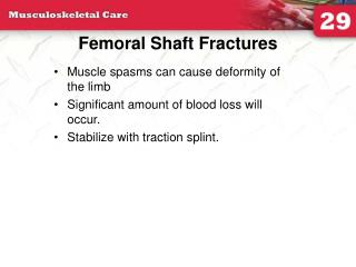
Initial Emerg Treatment • Gross Deformity Reduced and Stabilized • Traction: • Thomas Splint or Donway Splint
Initial Emerg Treatment • Traction cont’d: • Skin/Buck’s Traction • Maximum weight of 10 lbs
Initial Emerg Treatment • Traction cont’d: • Balanced Skeletal Traction • Unstable patient awaiting fixation • Helpful in keeping length • Historically used for definitive treatment • Distal femur vs. Tibia pin site
Classification: Winquist-Hansen • Type 0 • No Comminution • Type 1 • Small butterfly fragment • Transverse/short oblique # • Type 2 • Large butterfly frag. less than 50% width of bone
Classification:Winquist-Hansen • Type 3 • Large butterfly fragment • <50% cortical contact • Type 4 • Segmental comminution • No contact • Type 5 • Segmental bone loss
Surgical Timing • Within 24 hours associated with • Lower incidence of ARDS • Lower thromboembolic events • Improved rehab and shorter length of stay • Exceptions may exist for severe chest and head injuries • Damage Control
Sugical Timing • Fat Emboli ARDS • Major Criteria • Resp Symptoms- dyspnea and hypoxia • Petechial Rash – reddish brown, non-palpable, over upper body and *axilla* • CNS – Agitated Delirium • Radiologic Disease • Minor Criteria • Tachycardic, oral and retinal hemorrohages. febrile, renal insufficiency, elevated ESR, jaundice, drop Hgb + platletss • One Major + 4 minor + microglobulinemia = Dx • Treatment – Supportive therapy, rigid fracture fixation within 24 hours will decrease risk of ARDS
Surgical Timing • Damage Control Orthopaedics • ? Increased m and M with early fixation • Trauma is initial inflammatory response • Immediate fixation is a “Second Hit” • May cause patient to decompensate • Mitigated with provisional fixation • External Fixators
Surgical Timing • Morshed, JBJS Am (2009). Delayed Internal Fixation of Femoral Shaft Fracture Reduces Mortality Among Patients with Multisystem Trauma • “Delayed repair of femoral shaft fracture beyondtwelve hours in patients with multisystem trauma, which mayallow time for appropriate resuscitation, reduces mortalityby approximately 50%.” • Need to Consider systemic condition of patient
Surgical Timing • Open Fracture • Emergent I and D • IM nail within 24 hours • Same as closed fx • May require multiple washouts and delayed wound closure • High Grade Open Injuries (eg. 3B) • Consider External fixator, serial debridements and delayed conversion to IM Nail • Infection rate 3-4% • Non-union rate comparable to closed injury
Surgical Timing • Ipsilateral Femoral Neck Fracture • Priority to neck fracture fixation to avoid AVN • Cannulated screws + plate ORIF • Cannulated screws + retrograde nail • DHS + regrograde nail • All above have increased OR times • Cephalomedullary Nail/Recon Nail (TAN, FAN) • May not allow adequate femoral neck reduction, or have displacement of neck fracture during nailing
Treatment Options • Surgical Techniques • Traction • External Fixation • Plate Fixation • Intramedullary Nailing • Gold Standard is Reamed Antegrade IM Nail
Treatment Options • Traction • Mostly of historical significance • Disadvantages: prolonged hospital stay, traction sores, pin site infection, joint stiffness, muscle atrophy • Used for temporization these days
Treatment Options • External Fixator • Indications • Unstable polytrauma patient • High Grade Open Fractures • Vascular Injuries to restore length • Pt. Transfer • Most commonly monolateral ex-fix
Treatment Options • Plate Fixation • Indications: • Ipsilateral femoral neck fracture • Very small canal not suitable for nailing • Vascular injury requiring dissection • Femoral implants blocking femoral canal • ? Medicaly unstable patient (ARDS, pneumonia, etc.) • Bosse. JBJS 1997. Patients with thoracic injury. No change in survival rate between IM nail and ORIF with plate.
Treatment Options • Plate Fixation • Disadvantages • Disrupts normal # healing with callus • Higher infection incidence • Higher non-union and hardware failure
Treatment Options • Intramedullary Nailing • Antegrade vs. Retrograde • Piriformis vs. Trochanteric entry point • Reaming vs. Non-Reaming
Treatment Options: IM Nail • Antegrade • Gold Standard
Treatment Options: IM Nail • Retrograde • Gaining popularity • Start Point: Intracondylar notch, apex of Blumensaat’s line, 1 cm anterior to PCL origin
Treatment Options: IM Nail • Retrograde • Indications • Obesity, Pregnancy • IpsilateralTibial shaft #, neck # • Traumatic knee arthrotomy • Bilateral femur # • Ipsilateral TKR
Treatment Options: IM Nail • Retrograde
Treatment Options: IM Nail • Piriformis • Posterior = loss of proximal fixation • Anterior = Risk of bursting proximal segment
Treatment Options: IM Nail • Piriformis
Treatment Options: IM Nail • Trochanteric • Start point not necessarily at tip • Tip varies wrt long axis of femur • True start point is just lateral to the long axis of the femur
Treatment Options: IM Nail • Trochanteric
Treatment Options: IM Nail • Piriformis Vs. Trochanteric • Stannard et al. Functional Outcome Following Intramedullary Nailing of the Femur A Prospective Randomized Comparison of PiriformisFossa and Greater Trochanteric Entry Portals. JBJS (2011) • Equal for measures of postoperative hip function, union rates, and complication rates. • Higher HO in Piriformis group
Treatment Options: IM Nail • Reamed • Ream with Sharp, fluted, narrow Reamers • Ream to 0.5 to 1 mm size past chatter at isthmus • Over ream by 1 to 1.5 mm • Full speed, no stopping • Ensure central guide pin to avoid eccentric reaming
Treatment Options: IM Nail • Reamed
Treatment Options: IM Nail • Reamed Vs. Unreamed • Xin et al. Reamed intramedullary nailing versus unreamedintramedullary nailing for shaft fracture of femur: a systematic literature review. Arch OTS (2011). • No evidence for any difference in mortality or ARDS
Treatment Options: IM Nail • Reamed vs. Unreamed • Tornetta P, Tiburzi D. Reamed versus nonreamedanterograde femoral nailing. J Orthop Trauma, 2000 • No difference in OR time, transfusions, or hypoxic episodes • Blood loss greater with reamed • Time to union less for reamed
Complications • Malunion • Angular • Not very common with shaft #’s • More common in Subtroch or Distal Femur Fractures • Corrected with interference fit of nail
Complications • Malunion • Rotational • Internal or External rotation • External seen more with lateral patient position • Increased rotational malalignment with night-time surgery as well as fracture comminution
Complications • Malunion • Assessing Rotational Malalignment • Align ASIS, Patella, 2nd Ray • Compare LT profile • Assess Cortical Step Sign • Difference in thickness • Assess Diameter Difference sign • Femur is somewhat oval shaped • Achieve Anatomic Reduction
Complications • Malunion • Rotational • Occurs frequently BUT rarely symptomatic • Most common abnormality is ER • Tornetta n=22 average malrotation 16° 12/22 had > 10° malrotation 10/12 were in ER NO diff supine/ # table • Pt usually compensate well by rotating pelvis • If symptomatic • remove screws and derotate (early) / osteotomy (late)
Complications • Nonunion • Rate < 10% • Persistent pain – presenting symptoms • Related to degree of soft tissue damage • R/O Infection, inadequate stability, distraction of fracture site • X-ray for diagnosis, may require CT to confirm • Treatment options • Dynamization – used for distracted fractures • Exchange Nailing – reaming and changing nail • Union rates range – 53% to 96% • ORIF with Plate and bone grafting
Complications • Infection • <1% • Early infection (<3 months) • I and D, Abx • Nail removal and Ex. Fix or Abx spacer • Chronic Infection • Nail removal, ream for debridement • Abx. For 6 weeks • Definitive tx. when infx cleared (WBC/ESR/CRP)
Complications • Heterotopic Ossification • ~25% • Rarely clinically significant • Increased with piriformis entry point • Pudendal Nerve Injury~ 10% • Traction table • Femoral Artery or Nerve Injury • Perc interlocking screws with retrorade nail
Complications • Hardware failure • More common with plating • Weakness • Quads and Abductors • Iatrogenic fractures • Anterior start point with antegrade nail • Failure to overream canal • Evaluate Femoral Neck
OITE 1 • A 29-year-old male sustained a mid-shaft femur fracture in a motor cycle accident. Even if asymptomatic, what additional radiographs must be obtained either preoperatively or intraoperatively before performing intramedullary nailing of the femoral shaft fracture? 1. ipsilateral foot/calcaneus2. ipsilateral hip3. contralateral hip4. ipsilateral tibia/fibula5. thoracic spine
OITE • 2. Ipsilateral Hip • Ipsilateralfemoral neck fractures are seen in 1-9% of femoral shaft fractures and the femoral neck must be properly imaged either preoperatively or intraoperatively in any patient with a femoral shaft fracture. Dedicated hip films, possibly including an internal rotation AP, should be obtained before entering the OR. Daffner et al reported that in 11 of 20 cases of combined femoral shaft and neck fractures, the initial preoperative radiographs did not demonstrate the femoral neck fracture. Intraoperative fluoroscopy should also be used to evaluate for a femoral neck fracture both before (to evaluate for unrecognized fx) and after (to evaluate for iatrogenic fx) IM nailing. Tornetta et al also describe using preoperative CT scans to evaluate for a femoral neck fracture and found that they were able to reduce the number of missed ipsilateral femoral neck fractures.