Download
1 / 24
240 likes | 336 Views
Learn about tracheostomy, a surgical procedure to create a direct airway to the trachea. Explore its anatomy, types of tubes, procedure steps, complications, and management. Presented by Dr. Abdussalam M. Jahan, ENT specialist from Misurata University. Visit www.zahrawi.ly for more information.

E N D
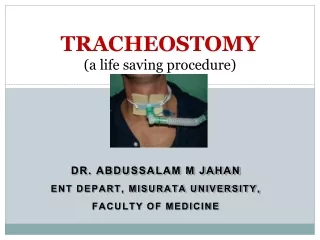
TRACHEOSTOMY(a life saving procedure) Dr. Abdussalam M jahan ENT depart, Misurata university, faculty of medicine
Introduction • Tracheostomy:is a surgical opening in the anterior aspect of the neck leading directly to the trachea. It is maintained open with tube called a Tracheostomy tube.
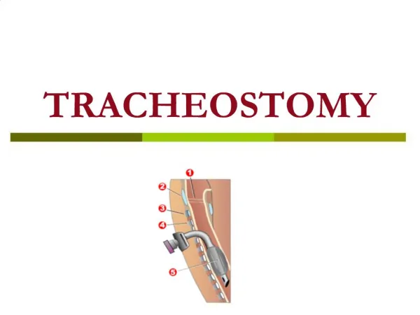
Relevant Surgical Anatomy 1 - Vocal cords 2 – Thyroid cartilage 3 - Cricoid cartilage 4 - Tracheal cartilages 5 - Balloon cuff
Indications: 1-upper airway obstruction, as mass or trauma, where intubation may be impossible. 2- Assist respiration over prolonged periods. 3-prolonged intubation with endotracheal tube: (more than 7-10 days) to reduce the possibility of subglottic stenosis.
4- assist cleaning of lower respiratory tract secretions 5-Facial fracturesthat may lead to upper airway obstruction (eg, comminuted fractures of the midface and mandible) 6-Major operations on the head and neck.
7- Reduce aspiration • 8-Severe obstructive sleep apnea. (if other options failed).
Contraindications • No absolute contraindications exist to tracheostomy • RELATIVE • Laryngeal carcinoma it may lead to increased incidence of stomal recurrence (adiffuse infiltrate of neoplastic tissue at the junction of the amputated trachea and skin )
Types of Tracheostomy Tubes Metal TT
Procedure • Transverse Incision • Incision 1 cm below the cricoid or halfway between the cricoid and the sternal notch.
Procedure cont’d • Blunt dissection of subcut tissue • Transversely retracted as shown
Procedure cont’d • Strap muscles are divided longitudinally at midline
Procedure cont’d • Thyroid ismuth is divided at midline by 2 hemostatic forcepses and cut edge secured by 2/0 vicryl
Procedure cont’d • After exposing of trachea, inverted U opening is made in trachea below 2nd ring
Procedure cont’d • By the Negus forceps the trachea dilated and tracheostomy tube inserted in the inverted U incision.
Procedure cont’d • Fixation of the tube.
Post-Op Management • Repeat X-Ray soft tissue neck • Strong Analgesia • Antibiotics • IV fluid until able to tolerate orally
Complications of Tracheostomy • Complications 5-40% • Mortality <2% • Complications are more frequent in emergency situations, severely ill patients
Immediate complications: • Apnea due to blood clot obstructing the tube, wrong insertion or displacement of the tube. • Bleeding. • Pneumothorax or pneumomediastinum: These can result from direct injury to the pleura or the cupola of the lung (especially in children). • Injury to adjacent structures: paratracheal structures vulnerable to injury are RLN, the great vessels, and the esophagus.
Early complications: • Early bleeding: This is usually the result of increased blood pressure as the patient recovery from anesthesia and begins to cough. Although this may necessitate a return to the operating room, bleeding may be controlled with local packing, cautery, control blood pressure. • Plugging with mucus.
Tracheitis: present in all patients with fresh tracheostomies. humidification, minimization of the fraction of inspired oxygen (FIO2) (because high oxygen levels exacerbate drying), and irrigation are essential. • Cellulitis. • Displacement of the tube.
Subcutaneous emphysema: This results from a tight closure of tissue around the tube, tight packing material around the tube, or false passage of the tube into pretracheal tissues. • Atelectasis: An overly long tube can mimic a unilateral mainstem intubation, causing atelectasis or collapse of the opposite lung.
Late complications: • Bleeding: Bleeding more than 48 hours after the procedure is most commonly (50%) caused by tracheo-innominate erosion (fistula). • Stenosis (subglottic stenosis) • Tracheoesophageal fistula: usually caused by friction between a posteriorly displaced tracheostomy tube or overinflated cuff and a rigid nasogastric tube. It manifests as aspiration and subsequent chemical pneumonia.
Tracheo-cutaneous fistula. • Scarring: Both vertical and horizontal incisions heal with small but visible scars. Clinico-Pathology Seminar: Dept. of ORL, UITH.