Download
1 / 53
530 likes | 551 Views
This text provides an overview of the functions and divisions of the nervous system, including how nerve impulses are generated and transmitted, the major structures of the central and peripheral nervous systems, and common diseases and disorders related to the nervous system.

E N D
Objectives: • Describe the functions of the nervous system • Identify and describe the functions of various types of nervous cells • Describe how a nerve impulse is generated and transmitted • Identify and describe the divisions of the nervous system • Identify the major structures of the central nervous system • Describe the major structure of the peripheral nervous system • List and describe diseases and disorders of the nervous system
Functions of the Nervous System • Sensory input – gathering information • To monitor changes occurring inside and outside the body • Changes = stimuli • Integration • To process and interpret sensory input and decide if action is needed • Motor output • A response to integrated stimuli • The response activates muscles or glands (effector organs)
Divisions of the Nervous System Structural classifications: • Central nervous system(CNS) • Brain • Spinal cord • Peripheral nervous system (PNS) • Nerve outside the brain and spinal cord • Sensory (afferent) and motor neurons (efferent)
Divisions of the Peripheral Nervous System Functional classifications: • Sensory (afferent) division • Nerve fibers that carry information to the central nervous system. Receptors of stimulus. • Motor (efferent) division • Nerve fibers that carry impulses away from the central nervous system to effector organs.
Divisions of the Efferent Peripheral Nervous System • Motor (efferent) division continued • Two subdivisions • Somatic nervous system = voluntary • Autonomic nervous system = involuntary • The Autonomic nervous system has two divisions: • Sympathetic = Becomes active when body is stressed, “fight of flight” • Parasympathetic = Becomes active when the body is relaxed or at rest.
Types of Neuroglial Cells and Their Functions • Microglia • Spider-like phagocytes • Dispose of debris • Ependymal cells • Line cavities of the brain and spinal cord • Circulate cerebrospinal fluid
Types of Neuroglial Cells and Their Functions • Oligodendrocytes • Produce myelin sheath around nerve fibers in the central nervous system • Satellite cells • Protect neuron cell bodies • Schwann cells • Form myelin sheath in the peripheral nervous system
Neuroglial Schwann Cells • Schwann cells – produce myelin sheaths in jelly-roll like fashion • Nodes of Ranvier – gaps in myelin sheath along the axon Figure 7.5
Neurons Neurons = nerve cells • Cells specialized to transmit messages • Major regions of neurons • Cell body – nucleus and metabolic center of the cell • Processes – fibers that extend from the cell body (dendrites and axon)
Neurons Classification of Neurons as it relates to their functions: • Sensory (afferent) neurons • Carry impulses from the sensory receptors • Cutaneous sense organs • Nerve endings (pain and temperature), Meissner’s corpuscle (touch), Pacinian corpuscle (deep pressure) • Proprioceptors – detect stretch or tension • Motor (efferent) neurons • Carry impulses from the central nervous system • Interneurons (association neurons) • Found in neural pathways in the central nervous system • Connect sensory and motor neurons
Neuron Structure • Cell body (Soma) • Nucleus • Large nucleolus • Nissl substance – specialized rough endoplasmic reticulum • Neurofibrils – intermediate cytoskeleton that maintains cell shape
Neuron Structure • Extensions outside the cell body • Dendrites – conduct impulses toward the cell body • Axons – conduct impulses away from the cell body • Axons end in axonal terminals • Axonal terminals contain vesicles with neurotransmitters (Ach) • Axonal terminals are separated from the next neuron by a gap • Synaptic cleft – gap between adjacent neurons • Synapse– junction between nerves The black arrows indicate the direction of the impulse movement along the neuron.
Pop Quiz Key Choices Oligodendrocytes Satellite Cells Schwann Cells Microglia Ependymal Cells Neuroglia Myelin Sheath 1. 3. 2. 4. 5. The type of cells in the diagrams are _____.
Pop Quiz Key Choices Node of Ranvier Axonal Terminal Dendrites Schwann Cell Microglia Soma 1. 2. 3. 4. 5.
Nerve Impulses • Irritability – ability to respond to stimuli • Conductivity – ability to transmit an impulse • The plasma membrane at rest is polarized • Fewer positive ions are inside the cell than outside the cell • resting potential difference is -70 millivolts between the outside and inside of the cell • inside of the cell’s axon contains K+ ions and Na+ ions are found outside of the axon’s membrane
Neuron Depolarization and Action Potentials • Depolarization– a stimulus depolarizes the neuron’s membrane • A deploarized membrane allows sodium (Na+) to flow inside the membrane through special proteins in the membrane called sodium channel proteins. • The movement of the ions initiates an action potential in the neuron due to the increase in voltage from -70 millivolts up to +30 millivolts within the axon • The action potential travels down the axon like a wave.
Neuron Depolarization and Action Potentials • If the action potential (nerve impulse) starts, it is propagated over the entire axon (all or none response) • When the axon’s internal charge reaches + 40 millivolts, the Na+ channels close and the K+ channels open; potassium ions rush out of the neuron after sodium ions have entered. The K+ ions move out until a negative charge of -70 millivolts is reestablished in the axon. Then the K+ channel proteins close. This repolarizes the axons membrane. However the Na+ and K + ions are in opposite locations of where they were before the neuron depolarized • The sodium-potassium pump restores the original configuration by pumping Na+ ions out and K + ions back into the axon. • This action requires ATP
Action Potentials and Neural Synapse • The impulse continues to move toward the cell body of the next neuron in the pathway. • Impulses are able to cross the synapse to another nerve • Neurotransmitter is released from a nerve’s axon terminal • The dendrite of the next neuron has receptors that are stimulated by the neurotransmitter • An action potential is started in the dendrite
Reflex Response • Reflex – rapid, predictable, and involuntary responses to stimuli • Reflex arc – direct route from a sensory neuron, to an interneuron, to an effector
Reflex Response • Autonomic reflexes – regulate involuntary activity • Somatic reflexes – all reflexes that stimulate skeletal muscles
Central Nervous System • CNS develops from the embryonic neural tube (ectoderm) • The neural tube becomes the brain and spinal cord • The opening of the neural tube becomes the ventricles • Four chambers within the brain • Filled with cerebrospinal fluid - nourishes and cushions the brain
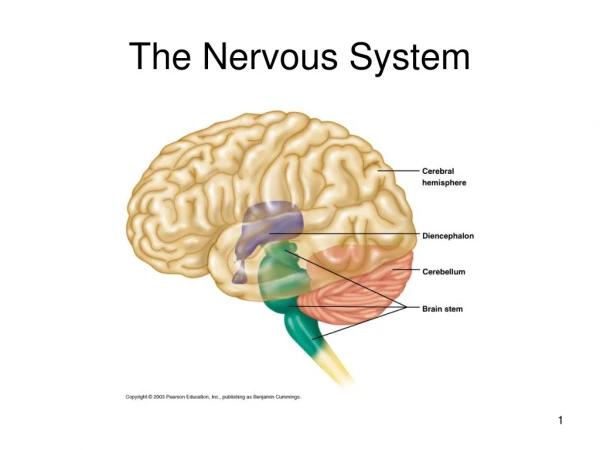
CNS: Brain: Cerebrum • Right hemisphere controls the left side of the body and the left hemisphere controls the right side of the body • more than half of the brain mass • surface is made of ridges (gyri) and grooves (sulci) • Fissures (deep grooves) divide the cerebrum into lobes • Surface lobes of the cerebrum • Frontal lobe • Parietal lobe • Occipital lobe • Temporal lobe
CNS: Brain: Diencephalon • Sits on top of the brain stem • Enclosed by the cerebral hemispheres • Made of three parts • Thalamus • Hypothalamus • Epithalamus
Diencephalon: Thalamus • relay station for sensory impulses; transfers impulses to the correct part of the cortex for localization and interpretation
Diencephalon: Hypothalamus • autonomic nervous system center; regulates body temperature, controls water balance, and regulates metabolism • an important part of the limbic system (emotions) and the pituitary gland is attached to the hypothalamus
Diencephalon: Epithalamus • houses the pineal body (an endocrine gland). • includes the choroid plexus which forms cerebrospinal fluid
CNS: Brain: Brain Stem • Attaches to the spinal cord • Parts of the brain stem • Midbrain • Pons • Medulla oblongata
Brainstem: Midbrain • composed of tracts of nerve fibers • two parts: cerebral peduncles and corpora quadrigemina • function - reflex centers for vision and hearing
Brainstem: Pons • mostly composed of fiber tracts • contains nuclei involved in the control of breathing
Brainstem: Medulla oblongata • lowest part of the brain stem • merges into the spinal cord • contains important control centers such as: heart rate control, blood pressure regulation, breathing, swallowing, and vomiting
CNS: Brain: Cerebellum • composed of two hemispheres with convoluted surfaces • provides involuntary coordination of body movements
specialized membranes below the bone surrounding the brain and spinal cord Dura mater (outer most layer) “tough mother” Double-layered external covering Periosteum – attached to surface of the skull Meningeal layer – outer covering of the brain Folds inward in several areas Arachnoid layer Middle layer Web-like Pia mater “gentle mother” Internal layer Clings to the surface of the brain Meninges
CSF: Cerebrospinal Fluid • Similar to blood plasma composition • Forms a watery cushion to protect the brain • Circulated in arachnoid space, ventricles, and central canal of the spinal cord
Nerve Anatomy • Nerve = bundle of neuron fibers • Neuron fibers are bundled by connective tissue • Endoneurium surrounds each fiber • Groups of fibers are bound into fascicles by perineurium • Fascicles are bound together by epineurium • Mixed nerves – both sensory and motor fibers
Quiz Key Choices Hypothalamus Cerebellum Diencephalon Cerebrum Thalamus Pons Brain Stem Medulla Oblongata 1. 2. 3. 4. 5. Which part of the brain helps you to vomit when you are sick? 6. Which part of the brain helps you to regulate body temperature? 7. Which part of the brain helps you to see and smell? 8. Which area of the brain includes the pons and medulla oblongata? 9. The hypothalamus is part of which area of the brain? 10. Which areas of the brain are divided into left and right hemispheres?
Diseases and Disorders of the Nervous System • Cerebrovascular Accident: CVA • Commonly called a stroke • The result of a ruptured blood vessel supplying (cerebral hemorrhage) a region of the brain or a vessel is obstructed by a clot. • Brain tissue supplied with oxygen from that blood source dies, swelling occurs in the brain due to leaking of blood from vessels. • Loss of some functions or death may result • This is due often to elevated blood pressure or hypertension.
Diseases and Disorders of the Nervous System Epilepsy: • This disease is due to random, mis-firing of neurons within the brain affecting sensory and motor regions of the brain. • Ranging in effects from sleep-like state of consciousness (narcolepsy), muscle paralysis and spasms (Petit mal and Grand mal seizures). Still not understood why this disease occurs. However in some cases it can result from brain trauma or injury.
Diseases and Disorders of the Nervous System • Concussion • Slight brain injury • No permanent brain damage • Contusion • Nervous tissue destruction occurs • Nervous tissue does not regenerate • Cerebral edema • Swelling from the inflammatory response or injury • May compress and kill brain tissue • May be caused by infectious agents such as viruses (encephalitis) or bacteria which cross the blood brain barrier or infect the meninges or CSF surrounding the brain (meningitis)


