Download
1 / 28
280 likes | 308 Views
Explore how sugar, fats, and proteins combine to produce waste products in the form of carbon dioxide, water, urea, and more. Learn about the functions of kidneys, anatomy of a nephron, urine formation process, components of urinalysis, and key pathological conditions affecting the urinary system.

E N D
WASTE PRODUCTS • Sugar and fats combined with oxygen (in the cells to produce energy) = carbon dioxide and water (as waste products). • Proteins combined with oxygen = nitrogenous wastes ENERGY SUGAR + CO2 + H2O FAT + OXYGEN
WASTE PRODUCTS • Nitrogenous Waste products: 1) Urea – formed in liver, travels in bloodstream to kidneys 2) Urine – travels down ureters into bladder and out of body 3) Creatinine 4) Uric acid
WASTE PRODUCTS • BUN = blood + urea + nitrogen • Measures urea levels in the blood. • Normally urea is excreted in urine, but if kidney is not functioning properly, then urea accumulates in the blood (Can lead to unconsciousness or death).
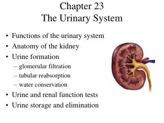
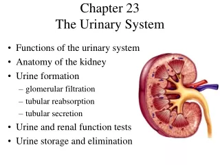
FUNCTIONS OF THE KIDNEYS • To remove urea & other nitrogenous wastes from the blood. • To maintain proper fluid & electrolyte balance in the body. • Forms urine • To act as an endocrine gland (by secreting): A) Renin – an enzymatic hormone that controls BP B) Vitamin D – needed to absorb calcium from intestine C) Erythropoetin – stimulates production of RBC’s in bone marrow.
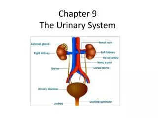
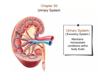
ORGANS INVOLVED • Kidneys – 2 bean shaped organs behind abdominal cavity (retroperitoneal), the outside region is called the cortex and the inside area is the medulla. • 2 Ureters – carry urine from kidney to bladder • Urinary Bladder – hollow sac in abdominal cavity that is a temporary reservoir for urine collection; trigone is area at base of bladder where ureters enter and urethra exits
Figure 7-5. Section of the kidney showing renal pelvis, calyces, & ureter.
ORGANS INVOLVED • Urethra – tube that carries urine from the bladder to outside of the body. • Micturation – voiding or elimination of urine
ANATOMY OF A NEPHRON • Nephron (glomerulus + renal tubule) • A million nephrons in each kidney. • Glomerulus – tiny ball of capillaries in cortex region of kidney. Glomeruli act as filter for water, salts, sugar & urea; a Bowman’s capsule surrounds each glomerulus & empties into renal tubule. (A) Renal artery branching to form smaller arteries, arterioles, and glomeruli. (B) Glomerulus & Bowman capsule. Afferent arteriole carries blood toward glomerulus. Efferent arteriole carries blood away from the glomerulus.
ANATOMY OF A NEPHRON 2) Proximal convoluted tubules – upper part of renal tubule 3) Loop of Henle – next part of renal tubule 4) Distal convoluted tubules – lower part of renal tubule 5) Collecting duct – portion of renal tubule that empties into central cavity of kidney (called renal pelvis).
URINE FORMATION • Urine formation – 3 processes: • Filtration – blood is filtered in the glomerulus allowing wastes (water, salt, sugar, urea, creatinine, uric acid) to leave bloodstream. Wastes collect in Bowman’s capsule. • Reabsorption – as wastes pass through the renal tubules, most of the water, some salt & all of sugars are reabsorbed into bloodstream. • Secretion – of some substances from bloodstream directly into renal tubule. These are waste products of metabolism (some acids, drugs, & potassium) that would become toxic if allowed to build-up in the body.
URINE FORMATION • Condensed version of urine formation:
COMPONENTS OF URINALYSIS • Urinalysis: examination of urine for abnormal substances • Color – yellow, amber or straw colored • Ph – normal 6.5 (slightly acidic) • Protein – small amount is normal, not normally detected; if protein is found, may indicate diabetes or hypertension • Glucose – not normally found • Specific Gravity – 1.010-1.025 normal, elevated in diabetes • Ketone Bodies – result from fat catabolism in cells (using fat for energy instead of sugar) • Sediment – abnormal particles in urine (WBC, RBC, bacteria, casts, etc…) • Pus – gives cloudy color to urine due to infection or inflammation • Bilirubin – results from hemoglobin breakdown
COMPONENTS OF URINE • Urine should not contain - sugar, albumin, blood, ketones, sediment, pus, or bilirubin • Urine should contain – water, urea, creatinine, salts, acids, drugs
COMBINING FORMS Cyst/o = urinary bladder cystocele = hernia of bladder Nephr/o = kidney nephro / lithiasis = kidney stone nephro / lithotomy = incision made to introduce scope to remove kidney stone Pyel/o = renal pelvis pyelo / lithotomy = incision made directly into kidney to remove kidney stone -tripsy = to crush lithotripsy = procedure to “crush” kidney stones Azot/o = nitrogen azotemia = nitrogen in blood (BUN) -uria = urination or glycosuria = sugar (glucose) in urine urine condition polyuria = excessive urination dysuria = painful or difficult urination
PATHOLOGICAL CONDITIONS • Glomerulonephritis – inflammation of kidney glomerulus; can be acute (post-strep) or chronic (HBP, albuminuria, renal failure, uremia, etc…) • Pyelonephritis – inflammation of renal pelvis and renal medulla; usually from a bacterial infection, abscesses form in kidney • Nephrotic Syndrome – group of symptoms caused by excessive protein loss (proteinuria, edema, hypoalbuminemia, hypercoagulability, hypercholesterolemia, and susceptible to infections) • Renal Failure – failure of kidney to excrete wastes and maintain filtration function
PATHOLOGICAL CONDITIONS • Diabetes Insipidus – inadequate secretion or resistance of kidney to action of ADH (antidiuretic hormone) Symptoms: • Dehydration – from excessive urination • Polydipsia – excessive thirst • polyuria – excessive urination • Diabetes Mellitus - inadequate secretion or improper utilization of insulin Symptoms: • Glycosuria – sugar in urine • Polydipsia – excessive thirst • Polyuria – excessive urination • Polyphagia – excessive hunger
ADD THESE DISORDERS TO YOUR NOTES! Acute cystitis. Notice that the mucosa of the bladder is red and swollen. Bladder and urinary tract infections are more common in women because of the shorter urethra, which allows easier bacterial colonization of the urinary bladder. They usually occur without a known cause, but may be acquired during sexual intercourse ("honeymoon cystitis") or following surgical procedures and urinary catheterization.
PATHOLOGICAL CONDITIONS Figure 7-8. Hydronephrosis caused by a stone (obstruction) in the proximal part of a ureter and hydroureter with hydronephrosis caused by a stone in the distal part of the ureter. Hydronephrosis is the swelling of the kidneys when urine flow is obstructed in any of part of the urinary tract. Swelling of the ureter, which always accompanies hydronephrosis, is called hydroureter. Hydronephrosis implies that a ureter and the renal pelvis (the connection of the ureter to the kidney) are overfilled with urine.
PATHOLOGICAL CONDITIONS Figure 7-9. (A) Polycystic kidney disease. The kidneys contain masses of cysts. Typically, polycystic kidneys weigh 20 times more than their usual weight (150-200 grams). (B) Chronic pyelonephritis. Notice that one kidney is small, shrunken, and irregularly scarred. The other kidney is of normal size but also shows scarring.
CLINICAL PROCEDURES • IVP (intravenous pyelogram) – Xray of kidney and ureters after injecting contrast dye into a vein • Cystoscopy – direct visual examination of urinary bladder with a scope. • Renal Angiogram – Xray of kidney blood vessels after injecting contrast dye. • VCUG (voiding cystourethrogram) – Xray of bladder, ureters, and urethra while pt. is voiding
CLINICAL PROCEDURES • Hemodialysis – artificial kidney machine that receives waste filled blood from patient, filters it, and returns filtered blood to patient. • Peritoneal Dialysis – fluid is instilled into peritoneal cavity through peritoneal catheter causing wastes to enter fluid; waste is removed when fluid drains.
CLINICAL PROCEDURES Continuous ambulatory peritoneal dialysis (CAPD). (A) The dialysis solution (dialysate) flows from a collapsible plastic bag through a catheter (a Tenckhoff peritoneal catheter) into the patient's peritoneal cavity. The empty bag is folded & inserted into undergarments. (B) After 4-8 hrs, the bag is unfolded, & the fluid is allowed to drain into it by gravity. The full bag is discarded, & a new bag of fresh dialysate is attached.
CLINICAL PROCEDURES • Ultrasonography – process of imaging urinary tract structures using high frequency sound waves • Renal Transplantation - surgical transfer of a complete kidney from a donor to a recipient
CLINICAL PROCEDURES Renal (kidney) transplantation. (A) Left kidney of donor is removed for transplant. (B) Kidney is transplanted to right pelvis of the recipient. The renal artery and vein of the donor kidney are joined to the recipient's artery and vein, & the lower end of the donor ureter is connected to the recipient's bladder (ureteroneocystostomy). The health of the donor is not affected by losing one kidney. In fact, the remaining kidney enlarges (hypertrophies) to take over almost full function.
CLINICAL PROCEDURESADD TO YOUR NOTES Figure 7-11. Cystoscopy.
CLINICAL PROCEDURES A Foley catheter in place in the urinary bladder.
ABBREVIATONS ADH: Antidiuretic Hormone ARF: Acute Renal Failure BUN: Blood Urea Nitrogen CI: Chloride (electrolyte excreted by kidney) CRF: Chronic Renal Failure HD: Hemodialysis IVP: Intravenous Pyleogram K+: Potassium (electrolyte) KUB: Kidneys, Ureters, Bladder Na+: Sodium (electrolyte) Sp gr: Specific Gravity UA: Urinalysis UTI: Urinary Tract Infection VCUG: Voiding Cystourethrogram