Download

1 / 11
110 likes | 138 Views
Explore the molecular basis of the genotype-phenotype relationship through DNA replication and polymerization as outlined in genetic analysis. Discover the factors influencing protein function and organism phenotype.

E N D
Chromosomal Landscapes From Figure 1-7 from Introduction to Genetic Analysis, Griffiths etal., 2012.
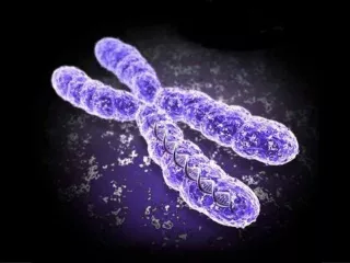
Human Chromosomal Landscapes From Figure 1-8 from Introduction to Genetic Analysis, Griffiths etal., 2012.
Molecular Basis for Relationship between Genotype and Phenotype genotype DNA DNA sequence transcription replication RNA translation amino acid sequence protein function phenotype organism
Replication of DNA is semiconservative. Each strand serves as a template. The two strands separate from each other when hydrogen bonds are broken. Replication of DNA is semiconservative. Each strand serves as a template. The two strands separate from each other when hydrogen bonds are broken. New strands are synthesized by the addition of nucleotides with bases complementary to those of the template. DNA replication is discontinuous. Two identical double helices result. New strands are synthesized by the addition of nucleotides with bases complementary to those of the template. DNA replication is discontinuous. Two identical double helices result. Refer to Figure 7-11 from Introduction to Genetic Analysis, Griffiths etal., 2012.
DNA polymerization requires DNA polymerase. Refer to Figure 7-15 from Introduction to Genetic Analysis, Griffiths etal., 2012.
DNA Polymerases • At least 5 DNA polymerases are known in E. coli . • DNA polymerase I (pol I): • adds nucleotides in 5’ to 3’ direction • removes mismatched bases in 3’ to 5’ direction • degrades double-stranded DNA in 5’ to 3’ direction • DNA polymerase II (pol II): • repairs interstrand cross-links • DNA polymerase III (pol III): • catalyzes DNA synthesis at replication fork in • 5’ to 3’ direction and only adds nucleotides at 3’ end of growing strand
Overview of DNA Synthesis DNA polymerases synthesize new strands in 5’ to 3’ direction. Primase makes RNA primer. Lagging strand DNA consists of Okazaki fragments. In E. coli, pol I fills in gaps in the lagging strand and removes RNA primer. Fragments are joined by DNA ligase.
DNA Replication at Growing Fork DNA polymerases add nucleotides in 5’ to 3’ direction. Because of antiparallel nature, synthesis of DNA is continuous for one strand and discontinuous for the other strand.
DNA Replication: Synthesis of Lagging Strand Several components and steps are involved in the discontinuous synthesis of the lagging strand. Note that DNA polymerases move in 3’ to 5’ direction on the template DNA sequence.
DNA Replication: Synthesis of Lagging Strand DNA extended from primers are called Okazakifragments. In E. coli, pol I removes RNA primers and fills in the gaps left in lagging strands. DNA ligase joins these pieces.