Download
1 / 58
620 likes | 886 Views
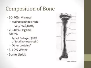
CHEMISTRY OF BONE. AND THE ROLE OF HORMONES IN MODELLING & REMODELLING By Dr Faiza Waseem. Bone. Inorganic (67%) Calcium hydroxyapatite Ca 10 (PO 4 ) 6 (OH) 2 along with sodium, magnesium, carbonate & fluoride. Organic (33%) component is called osteoid Type I collagen (28%)

E N D
CHEMISTRY OF BONE AND THE ROLE OF HORMONES IN MODELLING & REMODELLING By Dr Faiza Waseem
Bone • Inorganic (67%) • Calcium hydroxyapatite Ca10(PO4)6(OH)2 • along with sodium, magnesium, carbonate & fluoride.
Organic (33%) component is called osteoid • Type I collagen (28%) • Type V collagen is also present in small amounts • Non-collagen structural proteins (5%) • Proteoglycans • Sialoproteins • Phosphoproteins • Bone specific proteins: osteocalcin, osteonectin • Growth factors and cytokines (Trace)
Bone undergoes continuous turnover or remodeling throughout life • About 20% of bone is undergoing remodeling at any one time • Bone remodelling consists of : • resorption • Followed by deposition of new bone tissue • So adapts to physical & hormonal signals.
The cells involved in bone resorption and deposition are • osteoclast • osteoblast/cyte
OSTEOCLASTS • Osteoclasts are multinucleated • Derived from pluripotent hematopoietic stem cells. • A ruffled border • ATPase expels protons across the ruffled border into the resorption area. • pH lowered to 4 or less • increases the solubility of hydroxy apatite
Demineralization occurs • Lysosomal acid proteases released that digest the matrix proteins
OSTEOBLASTS • Mononuclear cells • Synthesize most of the proteins found in bone, growth factors and cytokines • Responsible for the deposition of new bone matrix (osteoid) & its mineralization • Control mineralization by regulating the passage of calcium and phosphate ions across their surface membranes. • The latter contain alkaline phosphatase which is used to generate phosphate ions from organic phosphates.
Alkaline phosphatase contributes to mineralization but in itself is not sufficient • Type 1 collagen is necessary • Acidic phosphoproteins eg bone sialoprotein act as sites of nucleation. • Contain poly-Asp and poly-Glu stretches that bind calciummineralization.
4% of compact bone and 20% of trabecular bone is renewed annually.
Factors affecting bone metabolism • PTH & VITAMIN D stimulate osteoblasts • Cortisteroids inhibit osteoblasts • PTH & VITAMIN D also stimulate osteoclasts • Calcitonin & estrogens inhibit osteoclasts.
Total calcium in the human body is 1-1.5 kg. • 99% present in bone • 1% in ECF • SOURCES: milk, cheese, egg yolk, nuts • The normal plasma calcium level=9-11mg/dl
Vitamin D • Vitamin D, after its activation to the hormone 1,25-dihydroxy Vitamin D3 is a principal regulator of Ca++.
Vitamin D is required for the intestinal absorption of calcium • Induces synthesis of calbindin • Vitamin D acts independently on bone • It increases the number and activity of osteoblasts, the bone forming cells. • Secretion of alkaline phosphatase by osteoblasts is increased by vitamin D
Synthesis of Vitamin D • Humans acquire vitamin D from two sources. • Vitamin D is produced in the skin by ultraviolet radiation and ingested in the diet.
Synthesis of Vitamin D • Vitamin D3 synthesis occurs in keratinocytes in the skin. • 7-dehydrocholesterol is photoconverted to previtamin D3, then spontaneously converts to vitamin D3. • Previtamin D3 will become degraded by over exposure to UV light and thus is not overproduced. • Also 1,25-dihydroxy-D (the end product of vitamin D synthesis) feeds back to inhibit its production.
Normal metabolism Vit D 25-HCC (Liver) Ca/PTH 1,25-DHCC 24,25-DHCC (Kidney) (Kidney)
Synthesis of Vitamin D • The mitochondrial P450 enzyme 1a-hydroxylase converts it to 1,25-dihydroxy-D, the most potent metabolite of Vitamin D. • The 1a-hydroxylase enzyme is the point of regulation of D synthesis. • Feedback regulation by 1,25-dihydroxy D inhibits this enzyme. • PTH stimulates 1a-hydroxylase and increases 1,25-dihydroxy D.
Synthesis of Vitamin D • Phosphate inhibits 1a-hydroxylase and decreased levels of PO4 stimulate 1a-hydroxylase activity • If excess 1,25-(OH)2-D is produced 24-hydroxylated to remove it.
Vitamin D (cholecalciferol) • Normal daily requirement • 200IU/day
Parathyroid Hormone • Secreted by parathyroid glands • Rapid response to reduced calcium (minutes) • Storage of PTH is only for about 1 hour.
Upon entry of pre-pro-PTH into endoplasmic reticulum, leader sequence is removed from the amino terminal to form the 90 a.a polypeptide pro-PTH. • Six additional a.a residues are removed from the amino terminal of pro PTH in the golgi apparatus and the 84 a.a polypeptide PTH is packaged in secretory granules and released as the main secretory product of the chief cells.
PTH action • The overall action of PTH is to increase plasma Ca++ levels and decrease plasma phosphate levels. • PTH acts directly on the bones to stimulate Ca++resorption and kidney to stimulate Ca++ reabsorption in the distal tubule of the kidney and to inhibit reabosorptioin of phosphate (thereby stimulating its excretion). • PTH also acts indirectly on intestine by stimulating 1,25-(OH)2-D synthesis.
Regulation of PTH Secretion and Biosynthesis • Extracellular Ca 2+ regulates secretion of PTH • Low Ca 2+ increases • High Ca 2+ decreases
An acute decrease of calcium marked increase of PTH mRNA increased rate of PTH secretion and synthesis. • However , 80-90% of the proPTH synthesized is quickly degraded. • This rate of degradation decreases when Ca+2 conc are low • & it increases when Ca+2 conc are high. • A Ca+2 receptor on the surface of the parathyroid cell mediates these effects.
Regulation of secretion • 1,25 dihydrocholecalciferol acts directlly on the parathyroid gland to decrease prepro PTH mRNA. • Increased plasma phosphate stimulate PTH secretion by lowering plasma Ca2+ and inhibiting the formation of 1,25 dihydroxy cholecalciferol. • Magnesium is required to maintain normal parathyroid secretory response .
Calcitonin • Secreted by the thyroid gland • Human calcitonin has M.W 3400 and contains 32 amino acid residues . • Effects are much less than those of PTH
Effects of Calcitonin • Decreases serum calcium level • Decreases the activity of osteoclasts and increases that of osteoblasts • Causes reduced bone turnover • PTH and Calcitonin are antagonistic. • Together they promote bone growth and remodelling. • In kidney calcitonin increases phosphorus excretion. This action is similar to PTH.
OSTEOPOROSIS • A generalized progressive reduction in bone tissue mass per unit volume. • The ratio of mineral to organic elements is unchanged in the remaining normal bone. • Skeletal weakness • Fractures • Estrogens & IL-1and IL-6 involved in the causation of osteoporosis.
Calcium, bones and osteoporosis • The total bone mass of humans peaks at 25-35 years of age. • Men have more bone mass than women. • A gradual decline occurs in both genders with aging, but women undergo an accelerated loss of bone due to increased resorption during perimenopause. • Bone resorption exceeds formation.
Calcium, bones and osteoporosis • Reduced risk: • Calcium in the diet • habitual exercise • avoidance of smoking and alcohol intake • avoid drinking carbonated soft drinks
Osteoporosis • Osteoporosis is the most common metabolic bone disease • WHO suggests that osteoporosis exists when bone density falls 2.5 standard deviations or more below the mean for young healthy adults of the same race and gender.
Factors that Affect Peak Bone Mass • Gender (M>F), males have greater PBM than females • Race (Blacks >Whites) • Genetics (osteoporosis runs in families and this may be the predominant factor) • Gonadal steroids (estrogen and testosterone increase bone mass) • Growth hormone (increases bone mass) • Calcium intake (supplements work) • Exercise (increases bone mass)
Primary osteoporosis • 3 types • Post-menopausal (Loss of oestrogen - incr osteoclastic activity).In males a decline in testosterone –incr osteoclastic activity) • Idiopathic (occurs in children and young adults) • Involutional (elderly)
Secondary Osteoporosis • uncommon • Nutrition - malnutr, malabs • Endocrine - Hyper PTH, Cush, • Drug induced - steroid, alcohol, smoking
Treatments • Exercise, activity • Calcium intake should be 1000-1500 mg/day • Males and females should take in 1000-1500 mg/day • All adults greater than 65 years should take 1500 mg/day • Three glasses of milk or three cups of yogurt per day provide 1000-1500 mg/day • Estrogen treatment • Estrogen inhibits osteoclastic activity • This therapy needs to be individualized • Estrogen may increase the incidence of breast cancer, heart attacks, stroke, blood clots
Treatments (Continued) • Bisphosphonates inhibit osteoclasts & are are classified as anti-resorptive medications • Calcitonin :Probably least effective Rx • Vitamin D • 400 IU per day
Rickets • It is due to deficiency of Vitamin D during childhood. The full-blown condition in children is characterized by weakness and bowing of weight bearing bones, dental defects and hypoclacemia.
OSTEOMALACIA • It is due to deficiency of vitamin D during adulthood, results from demineralization of bones, especially in women who have little exposure to sunlight.