Download
1 / 45
450 likes | 566 Views
Learn about the role of circulation and gas exchange in animals, from invertebrates to mammals. Explore the different types of circulatory systems, heart structures, blood pathways, and factors affecting heart rate. Discover the intricate mechanisms that maintain the beat and regulate blood pressure. Dive into the importance of arteries, veins, and capillaries in facilitating nutrient exchange and gas transfer. Enhance your understanding of the cardiac cycle and blood pressure dynamics in living organisms.

E N D
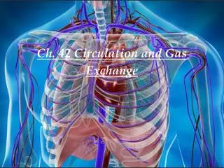
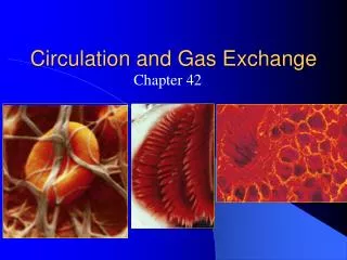
Circulation in Animals • Transport systems functionally connect the organs of exchange with body cells. • Diffusion is too slow. • By rapidly transporting fluid in bulk it connects the aqueous environment of cells to organs of gas exchange. • Nutrients are absorbed and wastes are taken away.
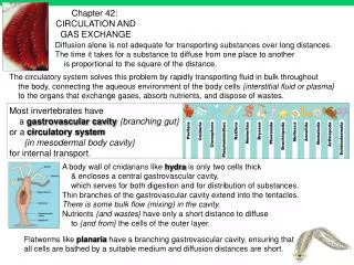
Invertebrate Nutrient and Gas Exchange • Gastrovascular Cavities • Simple organisms with few layers absorb nutrients through pockets in their bodies. • Extra-cellular digestion occurs to break down larger particles. • Cnidarians, Planarians, and Porifera • Circulation unnecessary because all cells are exposed to the environment.
Open Circulatory Systems • Open circulatory system • Blood bathes organs directly • Arthropods and most mollusks • No distinction between blood and interstitial fluid. • Body fluid called hemolymph. • Sinuses are spaces around organs. • One or more hearts pull blood in through ostia. • Blood squeezed around the body through movement.
Closed Circulatory System • Blood is confined to vessel and distinct from interstitial fluid. • Earth worms and higher animals. • Vertebrates have a cardiovascular system. • Atrium – top chamber of the heart • Ventricle- lower chamber of the heart. • Arteries – direct blood away from the heart. • Veins direct blood to the heart. • Capillary beds infiltrate each tissue.
Route of Blood Through the Body • Systemic circuit – delivers blood to the body. • Pulmonary circuit – picks up blood from pulmonary organs and delivers it back to the heart.
Fish Heart • Fish • Two chambers • Single circulation which means blood must travel through two capillary beds before it returns to the heart. • Because pressure drops in the capillary bed, blood flow is much slower than in other organisms.
AmphibianHeart • Three chambers • Two atria and one ventricle • A ridge separates most of the oxygenated from the deoxygenated blood, however some mixing does occur. • Double circulation • Blood returns to the heart after picking up oxygen. Sustains a higher pressure. • Pulmocutaneous circuit. • Pulmonary organs are the lungs and skin. • Reptiles • Three chambers clearly divided • Have a pulmonary circuit
Mammalian Heart • Two atria and two ventricles • Distinct separation of oxygen and deoxygenated blood. • Double circulation with a pulmonary circuit.
Mammalian Cardiac Cycle • Cardiac Cycle - One complete pumping and filling. • Systole – contraction phase of the cycle • Diastole – relaxation phase of the cycle. • Cardiac Output – the volume of blood that the left ventricle pumps into the systemic circuit per minute. • Depends on two factors: • Heart rate - rate of heart beat. • Stroke volume – blood pumped from left ventricle per contraction.
Average stroke volume for humans is 75 ml with a cardiac output of about 5.25 L/min. and an average pulse of 70 beats per minute. • Exercising increases stroke volume because the heart beats more efficiently. • More blood pumped to the systemic circuit per contraction.
The “Lub Dub” • Atrioventricular valves • Separate atria from ventricles. • Semilunar valves • Present at both exits of the heart. • Heart Murmur • Defect in the valves. • “Lub” • Recoil of blood against closed atrioventricular valves. • “Dub” • Recoil of blood against closed semilunar valves
Maintaining the Beat • Sinoatrial Node • Located in the right atrium near the Superior Vena Cava. • Sets the tempo for contraction. • Stimulates atrium to contract in unison. • Orchestration of muscle cells accomplished through intercalated discs. • Perkunji Fibers • Transmit electrical signal. • 0.1 second delay allows atria to completely empty before the ventricles contract
Maintaining the Beat • Atriovenricular Node • Causes the ventricles to contract. • Factors That Affect Heart Rate • Body temperature • 1o F increase increases heart rate by 10 beats/min. • Exercise • Hormonal stimulation. • Epinephrine –speeds up heart rate.
Arteries • Thicker walls • Outer wall connective tissue with elastic fibers • Middle layer smooth muscle – peristalsis • Inner wall – Endothelium , composed of smooth flat cells to reduce resistance to blood flow.
Veins • Thinner than arteries. • Valves to prevent back flow. • Blood moves through by muscle contractions. • Virtually all pressure from ventricular contraction is gone.
Capillaries • Lack outer and middle layers. • Basement membrane with endothelial layer. • Designed for gas and nutrient exchange between blood and interstitial fluid. • Blood flows 1000 times slower than in arteries and veins due to a large surface area. • Facilitates gas exchange. • Pressure drops dramatically in capillary beds
Blood Pressure • Hydrostatic pressure pushes blood through blood vessels. • Systole – pressure due to ventricular contraction. • Arteries stretch because blood flows out much more quickly than it can leave the artery. • Diastole – pressure due to the arterial wall ”snapping back”. • Next contraction occurs before all blood has left the artery and maintains pressure flow.
Blood pressure determined by: • Cardiac output • Peripheral resistance • Contraction of atrioles creates peripheral resistance. • Contractions triggered by stress, hormones, and nicotine. • Exercise increases cardiac output and dilates atrioles to supply blood to the muscles. • Giraffes and probably dinosaurs have valves and sinuses in their brain and neck to prevent a stroke while bending down for a drink.
Gas Exchange in the Capillaries • O2 and CO2 diffuse down their gradient. • Hydrostatic pressure on the atriole end forces 80% of the fluid out into the tissues. • Large proteins and blood cells left behind increases the osmolarity of the blood. • Causes fluid to move back into the capillaries at the venule end. • Fluid that doesn’t make it back into the cappilarie(15%) is returned to the blood through the lymphatic system.
Lymph Vessels • Lymph fluid composition about the same as interstitial fluid. • Moves through the body by squeezing of skeletal muscles and peristalsis. • Valves prevent back flow. • Lymph returns fluid that has leaked out in capillary beds to the circulatory system near the right atrium. • Lymph contains white cells that attack bacteria and viruses.
Plasma • Composition of Plasma • 90% H2O, inorganic salts (electrolytes) • PH about 7.4 • Plasma proteins • Buffers • Fibrinogen (clotting) • Proteins that escort lipids which are insoluble in water. • Hormones • Viscosity proteins • Kidneys maintain electrolytes.
Erythrocytes • Biconcave for more surface area. • Lack mitochondria and nuclei – more room for hemoglobin. • molecules of hemoglobin per cell. • Generate ATP through anaerobic metabolism. • Kidneys produce erythropoietin to stimulate the function of Red blood cells. • Hemoglobin also binds NO to relax capillaries.
Blood Pressure • Hydrostatic pressure pushes blood through blood vessels. • Highest during ventricular contraction. • During systole blood is pushed out into the artery faster than it can leave the artery. • During diastole artery “snaps back” and creates residual pressure in the arteries to continue pushing blood through. • Blood pressure is determined by cardiac output and peripheral resistance. • Stress contracts blood vessels and raises blood pressure. • Exercise causes atrioles to dilate causing more blood to flow in. A drop in pressure occurs and the cardiac output increases to compensate.
Cardiovascular Disease • Thrombus – blood clot. • Embolus – when clot breaks off and moves elsewhere in the body. • When a clot blocks a coronary artery – myocardial infarction. • Atherosclerosis – plaques form on the inner wall of the artery. • Arteriosclerosis – when plaques harden through calcification. • Angina Pectoris – heart pain that signals that the heart is not getting enough oxygen.
Hypertension • High Blood Pressure • Causes chronic abuse of the endothelium and encourages plaque formation. • Narrows the lumen of the blood vessels and reduces elasticity of blood vessels. • Can compromise arterial walls and increase risk for embolisms and aneurisms.