Circulation & Gas exchange
Circulation & Gas exchange. This will take your breath away…. Overview: Trading Places. Every organism must exchange materials with its environment Exchanges ultimately occur at the cellular level In unicellular organisms, these exchanges occur directly with the environment

Circulation & Gas exchange
E N D
Presentation Transcript
Circulation &Gas exchange This will take your breath away…
Overview: Trading Places • Every organism must exchange materials with its environment • Exchanges ultimately occur at the cellular level • In unicellular organisms, these exchanges occur directly with the environment • For most cells making up multicellular organisms, direct exchange with the environment is not possible • Gills are an example of a specialized exchange system in animals • Internal transport and gas exchange are functionally related in most animals
Open vs closed • In insects, other arthropods, and most molluscs, blood bathes the organs directly in an open circulatory system • In an open circulatory system, there is no distinction between blood and interstitial fluid, and this general body fluid is more correctly called hemolymph • In a closed circulatory system, blood is confined to vessels and is distinct from the interstitial fluid • Closed systems are more efficient at transporting circulatory fluids to tissues and cells
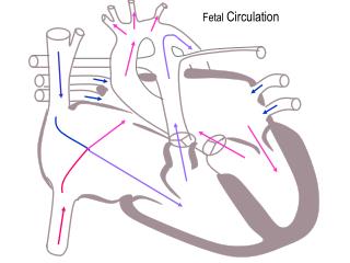
Mammalian Circulation • Blood begins its flow with the right ventricle pumping blood to the lungs • In the lungs, the blood loads O2 and unloads CO2 • Oxygen-rich blood from the lungs enters the heart at the left atrium and is pumped through the aorta to the body tissues by the left ventricle • The aorta provides blood to the heart through the coronary arteries • Blood returns to the heart through the superior vena cava (blood from head, neck, and forelimbs) and inferior vena cava (blood from trunk and hind limbs) • The superior vena cava and inferior vena cava flow into the right atrium
Fig. 42-6 Capillaries of head and forelimbs Superior vena cava 7 Pulmonary artery Pulmonary artery Capillaries of right lung Aorta 9 Capillaries of left lung 3 3 2 4 11 Pulmonary vein Pulmonary vein 5 1 Right atrium Left atrium 10 Right ventricle Left ventricle Inferior vena cava Aorta Capillaries of abdominal organs and hind limbs 8
Fig. 42-7 Pulmonary artery Aorta Pulmonary artery Right atrium Left atrium Semilunar valve Semilunar valve Atrioventricular valve Atrioventricular valve Right ventricle Left ventricle
The heart contracts and relaxes in a rhythmic cycle called the cardiac cycle • The contraction, or pumping, phase is called systole • The relaxation, or filling, phase is called diastole
The heart rate, also called the pulse, is the number of beats per minute • The stroke volume is the amount of blood pumped in a single contraction • The cardiac output is the volume of blood pumped into the systemic circulation per minute and depends on both the heart rate and stroke volume
Fig. 42-10 Artery Vein SEM Valve 100 µm Basal lamina Endothelium Endothelium Smooth muscle Smooth muscle Connective tissue Connective tissue Capillary Artery Vein Arteriole Venule 15 µm Red blood cell Capillary LM
Changes in Blood Pressure During the Cardiac Cycle • Systolic pressure is the pressure in the arteries during ventricular systole; it is the highest pressure in the arteries • Diastolic pressure is the pressure in the arteries during diastole; it is lower than systolic pressure • A pulse is the rhythmic bulging of artery walls with each heartbeat • Blood pressure is determined by cardiac output and peripheral resistance due to constriction of arterioles • Vasoconstriction is the contraction of smooth muscle in arteriole walls; it increases blood pressure • Vasodilationis the relaxation of smooth muscles in the arterioles; it causes blood pressure to fall
Fig. 42-16a Body tissue INTERSTITIAL FLUID Capillary Net fluid movement out Net fluid movement in Direction of blood flow
Fluid Return by the Lymphatic System • The lymphatic system returns fluid that leaks out in the capillary beds • This system aids in body defense • Fluid, called lymph, reenters the circulation directly at the venous end of the capillary bed and indirectly through the lymphatic system • The lymphatic system drains into veins in the neck • Lymph nodes are organs that filter lymph and play an important role in the body’s defense • Edema is swelling caused by disruptions in the flow of lymph
Fig. 42-17 Plasma 55% Constituent Major functions Cellular elements 45% Cell type Number per µL (mm3) of blood Functions Solvent for carrying other substances Water Erythrocytes (red blood cells) Transport oxygen and help transport carbon dioxide 5–6 million Ions (blood electrolytes) Sodium Potassium Calcium Magnesium Chloride Bicarbonate Osmotic balance, pH buffering, and regulation of membrane permeability Separated blood elements Leukocytes (white blood cells) Defense and immunity 5,000–10,000 Plasma proteins Albumin Osmotic balance pH buffering Lymphocyte Basophil Fibrinogen Clotting Eosinophil Immunoglobulins (antibodies) Defense Neutrophil Monocyte Substances transported by blood Nutrients (such as glucose, fatty acids, vitamins) Waste products of metabolism Respiratory gases (O2 and CO2) Hormones 250,000– 400,000 Platelets Blood clotting
Plasma • Blood plasma is about 90% water • Among its solutes are inorganic salts in the form of dissolved ions, sometimes called electrolytes • Another important class of solutes is the plasma proteins, which influence blood pH, osmotic pressure, and viscosity • Various plasma proteins function in lipid transport, immunity, and blood clotting
Erythrocytes • Red blood cells, or erythrocytes, are by far the most numerous blood cells • They transport oxygen throughout the body • They contain hemoglobin, the iron-containing protein that transports oxygen
Leukocytes • There are five major types of white blood cells, or leukocytes: monocytes, neutrophils, basophils, eosinophils, and lymphocytes • They function in defense by phagocytizing bacteria and debris or by producing antibodies • They are found both in and outside of the circulatory system
Platelets • Platelets are fragments of cells and function in blood clotting
Blood Clotting • When the endothelium of a blood vessel is damaged, the clotting mechanism begins • A cascade of complex reactions converts fibrinogen to fibrin, forming a clot • A blood clot formed within a blood vessel is called a thrombus and can block blood flow
Fig. 42-18-4 Red blood cell Collagen fibers Platelet plug Fibrin clot Platelet releases chemicals that make nearby platelets sticky Clotting factors from: Platelets Damaged cells Plasma (factors include calcium, vitamin K) Prothrombin Thrombin Fibrinogen Fibrin 5 µm
Fig. 42-22 Fluid flow through gill filament Oxygen-poor blood Anatomy of gills Oxygen-rich blood Gill arch Lamella Gill arch Gill filament organization Blood vessels Water flow Operculum Water flow between lamellae Blood flow through capillaries in lamella Countercurrent exchange PO2 (mm Hg) in water 150 120 90 60 30 Gill filaments Net diffu- sion of O2 from water to blood 110 80 50 20 140 PO2 (mm Hg) in blood
Fig. 42-24 Branch of pulmonary vein (oxygen-rich blood) Branch of pulmonary artery (oxygen-poor blood) Terminal bronchiole Nasal cavity Pharynx Larynx Alveoli (Esophagus) Left lung Trachea Right lung Bronchus Bronchiole Diaphragm Heart SEM Colorized SEM 50 µm 50 µm
Fig. 42-25 Rib cage expands as rib muscles contract Rib cage gets smaller as rib muscles relax Air inhaled Air exhaled Lung Diaphragm INHALATION Diaphragm contracts (moves down) EXHALATION Diaphragm relaxes (moves up)
Fig. 42-27 Cerebrospinal fluid Pons Breathing control centers Medulla oblongata Carotid arteries Aorta Diaphragm Rib muscles
Fig. 42-28 Alveolus Alveolus PCO2 = 40 mm Hg PO2 = 100 mm Hg PO2 = 40 PCO2 = 46 PCO2 = 40 PO2 = 100 Circulatory system Circulatory system PO2 = 40 PO2 = 100 PCO2 = 40 PCO2 = 46 PO2 ≤ 40 mm Hg PCO2 ≥ 46 mm Hg Body tissue Body tissue (a) Oxygen (b) Carbon dioxide