Download
1 / 17
170 likes | 268 Views
Embark on a captivating journey through the cell's framework, uncovering the intricate world of the cytoskeleton and protein synthesis. Discover the vital role of microfilaments, intermediate filaments, and microtubules, and witness organelles like the Endoplasmic Reticulum and Golgi apparatus in action. Unravel the process of protein translation, from mRNA transcription to tRNA's role in building proteins within ribosomes. Explore the fascinating genetic code that encodes amino acids and learn about the essential components of tRNA and ribosomes in protein synthesis. Dive deep into the cellular processes that shape life as we know it.

E N D
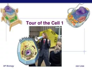
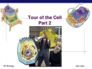
A tour through the Cell cont… The framework of a cell - the Cytoskeleton Organelles in action - Protein Synthesis
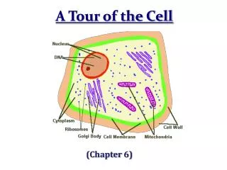
Cytoplasm • semi-fluid-like jelly within the cell • division into three subdivisions: cytosol, cytoskeleton & organelles
Cytoskeleton: • internal framework of the cell • gives the cytoplasm flexibility and strength • provides the cell with mechanical support • gives the cell its shape • can be rapidly disassembled in one area of the cell and reassembled in another • anchorage points for organelles and cytoplasmic enzymes • also plays a role in cell migration and movement by the cell
Cytoskeleton: • three major components • 1. microfilaments • 2. intermediate filaments • 3. microtubules
microfilaments = thin filaments (7 nm) made up of a protein called actin -twisted double chain of actin subunits -forms a dense network immediately under the PM (called the cortex) -also found scattered throughout the cytoplasm -function: 1.anchor to membrane proteins 2. interaction with myosin = interacts with larger microfilaments made up of myosin - results in active movements within a cell (e.g. muscle cell contraction) 3. provide much of the mechanical strength of the cell 4. give the cell its shape 5. also provide support for cellular extensions called microvilli (small intestines)
intermediate filaments = range from 8 to 12 nm in diameter -function: 1. impart strength to the cytoskeleton (like microfilaments) 2. support cell shape 3. anchors & stabilize organelles 4. transport materials within a cell
microtubules= hollow rods or “straws” of 25 nm in diameter - made of repeating units of proteins called tubulin - function: 1. cell shape & strength 2. organelles: anchorage & movement 3. mitosis - form the spindle(chromosome movement) 4. form many of the non-membranous organelles - cilia, flagella, centrioles
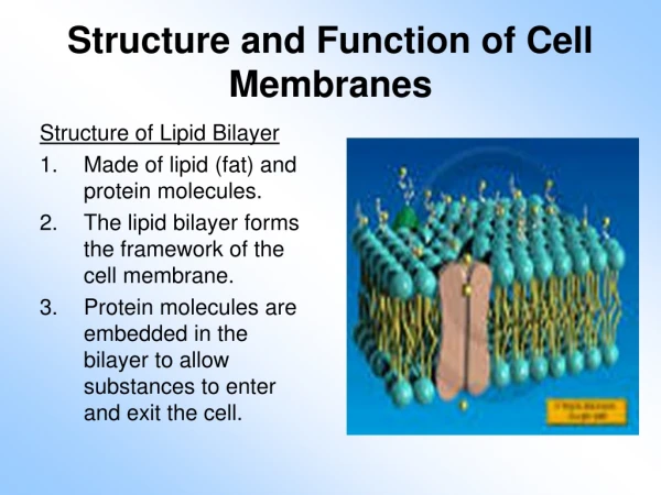
Organelles in Action • organelles attach to the cytoskeleton – held in place • each organelle has a distinct function • organelle of protein synthesis = Endoplasmic Reticulum • large organelle surrounded by a phospholipid bilayer and attached to the nucleus • can be found studded with ribosomes = Rough ER (protein synthesis) • parts found without ribosomes and make lipids = Smooth ER • organelle of protein modification and packaging = Golgi apparatus
Protein synthesis • known as translation • translating the message found in DNA/RNA into a polypeptide chain protein • requires three things • 1. mRNA – messenger RNA transcribed from the DNA template • 2. tRNA – transfer RNA that carries the amino acids of the future protein • 3. ribosome – the “machine” of translation
How are the instructions for assembling amino acids into proteins encoded in your DNA? • first the DNA gets transcribed into a message = mRNA • the mRNA gets exported out into the cytoplasm • the mRNA gets bound by a ribosome • tRNA molecules bring the correct amino acid into the ribosome • amino acids are linked together mRNA
Protein Translation: The Genetic Code Second mRNA base A U C G UUU UAU UCU UGU U Phe Cys Tyr UUC UCC UAC UGC C U Ser UUA UCA UGA Stop A UAA Stop Leu • the mRNA nucleotide sequence is “read” by the ribosome in groups of 3 nucleotides = “codon” • each codon codes for 1 of the 20 amino acids that make up proteins in eukaryotes • all of these codons grouped together is called the “genetic code” • the code is redundant - each amino acid can be coded for by more than one codon • e.g. alanine – GCU, GCC, GCA and GCG • the GC defines the amino acid as alanine • in many cases the 3rd codon is important in defining the amino acid • serine – codons are: AGU, AGC • BUT arginine codons are: AGA and AGG Trp UUG UCG UGG G UAG Stop CUU CCU U CAU CGU His CUC CAC CGC C CCC C Leu Pro Arg CUA CCA CGA A CAA Gln CUG CCG CGG G CAG First mRNA base (5 end of codon) Third mRNA base (3 end of codon) AUU AAU ACU AGU U Ser Asn C AUC Ile AAC ACC AGC A Thr AUA AAA ACA AGA A Lys Arg Met orstart AUG ACG AGG AAG G GUU GCU GAU GGU U Asp GUC GCC C GGC GAC G Val Ala Gly Gly GUA GCA GGA A GAA Glu GUG GCG GGG G GAG
Building a protein: tRNA • where do the amino acids come from • they are brought into the ribosome bound to tRNA molecules • tRNA molecule consists of a single strand of RNA - about 80 RNA nucleotides long • at one end – anticodon site for binding with the mRNA template • at the other end – attachment site for the amino acid that corresponds to the mRNA codon • 3 • Amino acidattachmentsite • 5 • Amino acidattachmentsite • 5 • 3 • Hydrogenbonds • Hydrogenbonds A A G • 3 • 5 • Anticodon • Anticodon • Anticodon (a) Two-dimensional structure (b) Three-dimensional structure (c) Symbol used in books
Building a Protein: Ribosomes • machine of translation • made in the nucleolus in eukaryotic cells • comprised of two subunits of proteins (large and small) linked together • eukaryotes: small subunit = ~33 proteins + large subunit = ~50 proteins • subunits are exported out via nuclear pores
Ribosomes Growing polypeptide Amino end • within the large subunit are two sites for the binding of tRNAs • P-site or Peptidyl-tRNA site – “old” AA • A-site or aminoacyl-tRNA site – incoming AA • and one E site/Exit site for the exit of the old tRNA off the ribosome Next aminoacid to beadded topolypeptidechain E tRNA mRNA 3 Codons 5 (c) Schematic model with mRNA and tRNA
Translation http://highered.mcgraw-hill.com/sites/0072507470/student_view0/chapter3/animation__how_translation_works.html
Organelles in Disease: The lysosome Lysosomes= “garbage disposals” -dismantle debris, eat foreign invaders/viruses taken in by endocytosis or phagocytosis -also destroy worn cellular parts from the cell itself and recycles the usable components = autophagy -form by the budding of vesicles off the Golgi and their fusion -acidic interior -1. contain enzymes that breakdown DNA, RNA (nucleases) and proteins (proteases) -2. contains enzymes for the breakdown of lipids and phospholipids TaySachs and lysosomes:human genetic disease -severe mental degradation -lysosomes lack one of the 40 required enzymes -results in a build up of fatty material on neurons -failure of nervous system communication -infantile form of the disease = death by 4 yrs -juvenile form = death from 5 to 15 yrs -adult onset – not fatal; progressive loss of nervous function -most common in Ashkenazi Jews, French Canadians and Cajun populations in Louisiana (same mutation as Jews)
Organelles in Disease: The Peroxisome -only identified in 1954 -found in all cells – abundant in liver and kidney cells -major function is breakdown of long chain fatty acids -other functions: 1. synthesis of bile acids 2. breakdown of alcohol by liver cells 3. anti-oxidant function - contains enzymes to break down dangerous chemicals made by the cell during metabolism Adrenoleukodystrophy and peroxisomes: -X linked disorder -1:20,000 to 1:50,000 births -peroxisomes can’t break down fatty acids properly -leads to a build up of big, saturated fatty acids on cells of throughout the body -can result in neuron death – not known why -lethargy, skin darkens, blood sugar drops, altered heart rhythm due to imbalanced electrolytes, paralysis, death *** slowed by a certain triglyceride found in rapeseed oil Lorenzo Odone = “Lorenzo’s Oil” (mixture of unsaturated fatty acids that slows the development of these saturated FAs) F-actin and peroxisomes