Download
1 / 28
350 likes | 956 Views
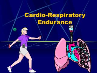
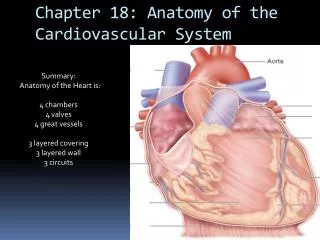
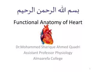
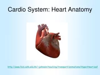
Cardio System: Heart Anatomy. http://www.fed.cuhk.edu.hk/~johnson/teaching/transport/animations/HyperHeart.swf. The Cardiovascular System. The Cardiovascular System consists of the heart and the blood vessels It is a closed system of blood vessels through

E N D
Cardio System: Heart Anatomy http://www.fed.cuhk.edu.hk/~johnson/teaching/transport/animations/HyperHeart.swf
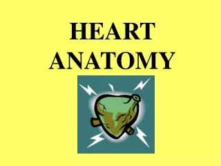
The Cardiovascular System • The Cardiovascular System consists of the heart and the blood vessels • It is a closed system of blood vessels through • which the blood, a fluid connective tissue, is • propelled by the heart, a muscular pump • The heart functions to pump blood through the • blood vessels to all body tissues, allowing for • transport of nutrients, oxygen, CO2, wastes, and • hormones
The Cardiovascular System The heart is located in the thoracic cavity, In the mediastinal space, between the lungs
The heart is divided into a right and a left side • Right Heart • Receives blood from the body • Contains deoxygenated blood • Will be pumped to the lungs to receive oxygen • Left Heart • Receives blood from the lungs • Contains oxygenated blood • Will be pumped to all body tissues
The Cardiovascular System There are three types of blood vessels • Arteries • Carry blood away from the heart • Veins • Return blood to the heart • Capillaries • Allow exchange of substances between the blood and extracellular fluid
Heart Anatomy External View • Base • Apex
Heart Anatomy External View The Pericardium is the protective covering over the heart. Heart is located in the pericardial cavity It has two functions: • Protection • Reduce Friction
Photo of the pericardium cut open to show the heart The pericardium protects the heart and also reduces friction as the heart beats The pericardium has 2 layers • Fibrous layer • Tough outer layer • Fibrous connective tissue • Serous layer • Delicate inner layer • Provides fluid for lubrication
Heart Anatomy • The pericardium has 2 layers • Fibrous Pericardium • Serous Pericardium • Parietal Layer • Pericardial Space • Visceral layer • (Epicardium)
The Heart Wall • The heart wall is made of three layers • Epicardium • Myocardium • Endocardium
The Heart Wall • There are three layers of the heart wall • Epicardium – outer layer (visceral pericardium) • Myocardium – middle layer • Makes up the majority of • the heart’s mass • Endocardium – inner layer • Lines the chambers, valves, vessels
The Heart Wall • There are three layers of the heart wall • Epicardium • Outer layer • Visceral pericardium • Myocardium • Middle layer • Makes up the majority of the heart’s mass • Endocardium • Inner layer • Lines the chambers, valves, vessels
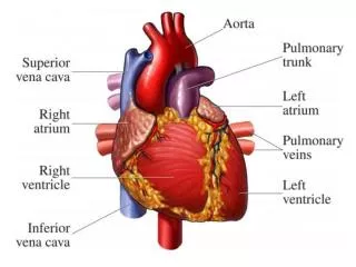
Heart Chambers There are 4 heart chambers • Right Atrium • Left Atrium • Right Ventricle • Left Ventricle
Heart Valves There are 4 heart valves • Right Atrioventricular (A-V) Valve • Tricuspid • Left Atrioventricular (A-V) Valve • Bicuspid, Mitral • Pulmonary Semilunar Valve • Aortic Semilunar Valve
Blood Vessels • Blood Vessels • Superior and Inferior Vena Cava • Pulmonary Trunk • Pulmonary Arteries • Pulmonary Veins • Aorta
Coronary Blood Vessels External View Coronary blood vessels supply the myocardium with blood • Coronary Arteries • First Branches off the Aorta • Coronary Veins • Return blood to the Cranial Vena Cava
Internal Anatomy Muscular divisions • Interatrial Septum • b/t atria; serves to separate artial bld • Interventricular Septum • b/t ventricles; serves to separate ventricular bld • Atrioventricular Septum • b/t ventricles; serves to separate artial bld and ventricle bld
Internal Anatomy • Chordae Tendinae • In ventricles; attach to mitral (bicuspid) and tricuspid valves and regulate opening and closing of valves. • Papillary Muscles • In ventricular wall; attach heart strings
Internal Anatomy Were you paying attention??? Identify: #9 #10 #11 #12 #13 #14 #15 #16 #17
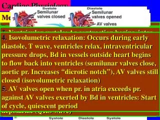
Blood Flow Through the Heart Blood enters the right and left atria Blood enters the ventricles Blood is pumped to the lungs and body
Blood Flow Through the Heart Blood returns to the heart from the cranial part of the body through the cranial vena cava, and from the caudal part of the body through the caudal vena cava,to the right atrium It then flows as follows: Right A-V Valve Right Ventricle Pulmonary Semilunar Valve Pulmonary Arteries (R & L) to the Lungs Pulmonary Veins (R & L) Left Atrium Left A-V Valve Left Ventricle Aortic Semilunar Valve Aorta to the body
http://www.nhlbi.nih.gov/health/dci/Diseases/hhw/hhw_pumping.htmlhttp://www.nhlbi.nih.gov/health/dci/Diseases/hhw/hhw_pumping.html
Heart Chambers http://www.heartpoint.com/theheart.html