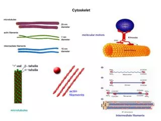
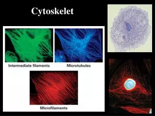
Cytoskelet
Cytoskelet. Tubulin jako GTP-áza. Samouspořádánà mikrotubulárnÃho cytoskeletu. Aktin a jeho polymerace. Stabilizace vs. destabilizace mikrotubulů, mikrotubuly vazebné proteiny. Aktin-vazebné proteiny. All myosins have a similar mechanochemical cycles.

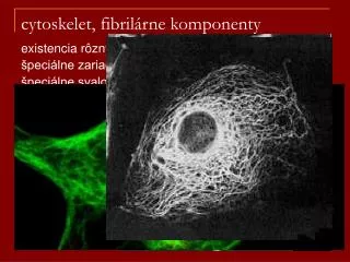
Cytoskelet
E N D
Presentation Transcript
Stabilizace vs. destabilizace mikrotubulů, mikrotubuly vazebné proteiny
All myosins have a similar mechanochemical cycles ATPase Cycle “The Movie” PG-13 So how do myosins differ?
Myosin V transports vesicles in cells • Myosin V is a “processive” motor YFP YFP 10 m Myosin V HMM
Myosin II is a low duty ratio (< 10%) motor Myosin V -long neck (6IQ motifs) -organelle motor -functional unit: two heads Myosin II -short neck (2IQ motifs) -drives muscle contraction -functional unit: ~20 heads
Many myosin II molecules required to propel actin filament Myosin 30 m In vitro motility assay Real time fluorescence microscopy
In muscle, myosin II molecules are assembled into a thick filament. • Filaments form by association of hydrophobic regions in the tail.
Muscle sarcomere is the fundamental contractile unit in muscle. • The sarcomere contracts when myosin thick filaments and actin thin filaments slide past each other.
Myofibril consists of a series of sarcomeres • Light and dark banding pattern gives rise to “striated” muscle. A band I band Figure 16-69 Molecular Biology of the Cell
Sarcomere shortening (i.e., muscle contraction) shortens the I band but not the A band.
“Crowns” of crossbridges project from thick filament at 14.3 nm intervals and successive crowns are rotated. • The result is a thick filament with six rows of crossbridges along its length.
Thick and thin filaments in an insect flight muscle Thick filament Myosin Thin filament Actin
Accessory proteins in muscle • CapZ and tropomodulin cap ends of actin to keep filament length constant. • Z disc contains -actinin and other proteins that stably join sarcomeres. • Titin maintains thick filament position in the sarcomere. • Nebulin sets the length of the thin filaments. Figure 16-72 Molecular Biology of the Cell
Muscle growth • Hypertrophy: Adding new myofibrils within a cell. • Hyperplasia: Formation of new cells. • Lengthening: Adding more sarcomeres in series.
Myosin mutations cause “Familial hypertrophic cardiomyopathy” and sudden death Mouse model of hypertrophic cardiomyopathy Wt cardiac myosin R403N mutant myosin