Download
1 / 1
10 likes | 160 Views
Retinal nerve fiber layer thickness change in patients with wet AMD treated with ranibizumab, short term results. Özen Osmanbaşoğlu, MD 1 Zeynep Alkın, MD 2 Ahmet Taylan Yazıcı , Ass. Prof. 2 Hülya Güngel, Prof. Dr 1 1.Istanbul Education and Research Hospital

E N D
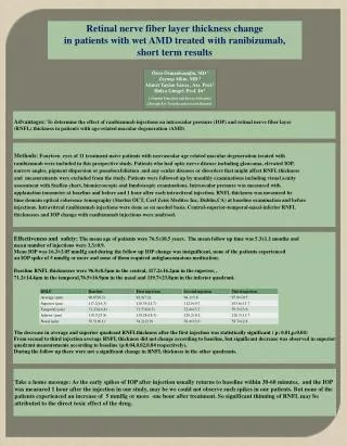
Retinal nerve fiber layer thickness change in patients with wet AMD treated with ranibizumab, short term results Özen Osmanbaşoğlu, MD 1 Zeynep Alkın, MD 2 Ahmet Taylan Yazıcı , Ass. Prof.2 Hülya Güngel, Prof. Dr1 1.Istanbul Education and Research Hospital 2.Beyoglu Eye Training and research Hospital Advantages: Todeterminetheeffect of ranibizumabinjections on intraocularpressure (IOP) andretinalnerve fiber layer (RNFL) thickness in patientswithage-relatedmaculardegeneration (AMD) Methods: Fourteen eyes of 11 treatment naive patients with neovascular age related macular degeneration treated with ranibizumab were included in thisprospectivestudy. Patientswho had opticnervediseaseincludingglaucoma, elevated IOP, narrowangles, pigment dispersionorpseudoexfoliationandanyoculardiseasesordisordersthatmightaffect RNFL thickness andmeasurementswereexcludedfromthestudy. Patients were followed up by monthly examinationsincludingvisualacuity assessmentwithSnellenchart, biomicroscopicandfundoscopicexaminations. Intraocular pressure was measuredwith applanationtonometerat baselineandbefore and 1 hour after eachintravitreal injection. RNFL thickness was measured by time domainoptical coherence tomography (Startus OCT, Carl ZeissMeditecInc, Dublin,CA)at baselineexaminationandbefore injections.Intravitrealranibizumab injections were done as on needed basis. Central-superior-temporal-nasal-inferior RNFL thicknesses and IOP changewithranibizumabinjections were analysed. Effectiveness and safety: The mean age of patients were 76.5±10.5 years. The mean follow up time was 5.3±1.1 months and mean number of injections were 3.3±0.9. Mean IOP was 16.2±2.05 mmHg and during the follow up IOP change was insignificant, none of the patients experienced an IOP spike of 5 mmHg or more and none of them required antiglaucomatous medication. Baseline RNFL thicknesses were 96.8±8.5µm in the central, 117.2±16.2µm in the superior, , 71.2±14.4µm in the temporal,76.5±16.9µm in the nasal and 119.7±23.8µm in the inferior quadrant. The decrease in average and superior quadrant RNFLthickness after the first injection was statistically significant ( p: 0.01,p:0.04) From second to third injection average RNFL thickness did not change according to baseline, but significant decrease was observed in superior quadrant measurements according to baseline (p:0.04,0.02,0.04 respectively). During the follow up there were not a significant change in RNFL thickness in the other quadrants. . Take a homemessage: As theearlyspikes of IOP afterinjectionusuallyreturnstobaselinewithin 30-60 minutes, andthe IOP wasmeasured 1 houraftertheinjection in ourstudy, may be wecould not observesuchspikes in ourpatients. But none of the patientsexperiencedan increaseof 5 mmHgormoreonehouraftertreatment. Sosignificantthinning of RNFL may be attributed to the direct toxic effect of the drug.