Knee Anatomy
540 likes | 717 Views
This detailed presentation on knee anatomy highlights the bony structures, ligaments, and joint mechanics essential for orthopaedic professionals. Key components such as the tibiofemoral joint, patellofemoral joint, menisci, and critical ligaments like the ACL and PCL are discussed in depth. Insights into the biomechanics of knee movement, joint reactive forces during activities, and significant anatomical relationships of nerves and arteries are explored. This knowledge enables better clinical understanding and enhanced surgical outcomes in knee reconstruction and sports medicine.

Knee Anatomy
E N D
Presentation Transcript
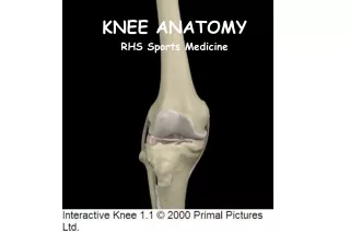
Knee Anatomy Reza Omid, M.D. Assistant Professor Orthopaedic Surgery Shoulder & Elbow Reconstruction Sports Medicine Keck School of Medicine of USC
Bony Anatomy • Tibiofemoral joint • Patellofemoral joint
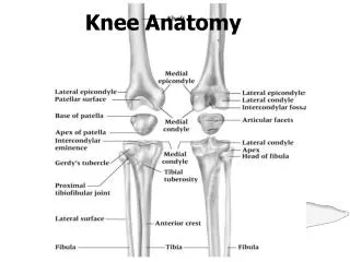
Femoral Condyles • A – Lateral Condyle • Smaller radius of curvature • Smaller in all dimensions • Extends more anteriorly • B – Medial Condyle • Larger radius of curvature • Extends more distally • C – Intercondylar notch
Tibial Plateau • D – Medial Plateau • Greater surface area • Concave • Circular shape • E – Intercondylar Eminence • F – Lateral Plateau • Smaller surface area • Convex • Oval shape
Patella • Sesamoid bone in quadriceps • Dividing central ridge • Comprised of seven facets • Medial and Lateral facets divided into 3rds • 7th facet is most medial (odd facet) • Medial half usually smaller • Thick hyaline cartilage (5.5mm)
Femoral Sulcus • Lateral wider and higher • Both with sagittal convexity
Screw Home Mechanism • Knee achieves terminal extension via the “screw home mechanism • The tibia externally rotates in relation to the femur. • When the knee needs to flex, the popliteus contracts which causes internal rotation of the tibia and in essence unlocking the knee and allowing it to bend
Popliteal Artery • Originates at the adductor hiatus and passes through the popliteal fossa, then deep to the fibrous arch over the soleus muscle • Divides into the anterior and posterior tibial arteries at the distal aspect of the popliteus muscle
Popliteal Artery • The popliteal artery is 9mm posterior to the posterior cortex of the tibia at 90° of flexion and even closer in extension. • Place retractors biased to the medial side when possible.
Skin Blood Flow • If two longitudinal incisions are present, the more lateral incision should be used (if allows adequate exposure) because most of blood supply comes in medially. • The lateral skin edge is more hypoxic than the medial skin edge so keep this in mind when placing sutures.
Tibial Nerve • Initially lateral to the popliteal artery • Crosses at midpoint to end medial to the artery at soleus arch
Common Peroneal Nerve • Lateral aspect of the popliteal space • Medial and posterior to the biceps femoris tendon
Patellofemoral Biomechanics • Joint Reactive Force • In flexion, patella compressed onto femur creating joint reactive force • Stair climbing – 3.5 X BW • Deep bends – 7-8 X BW
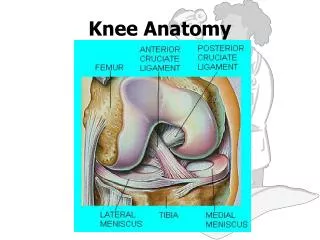
Menisci • Primarily type I collagen with fibers arranged obliquely, radially, and vertically • Outer 10% to 30% has blood supplied from the perimeniscal capillary plexus off the superior and inferior medial and lateral genicular arteries
Meniscus Function • Load Transmission • 50% load transmitted in extension • 85% load transmitted at 90 degrees flexion • Resection of 15-34% increases pressure 350% • Secondary Stabilization • Medial meniscus provides anterior restraint • Especially in ACL deficient knee
Lateral Meniscus • Loose peripheral attachment allows greater translation during motion • Average excursions of the menisci with knee flexion • 5.2 mm for the medial • 11 mm for the lateral • Bare area anterior to popliteus tendon • Two highly variable meniscofemoral ligaments attach it to medial femoral condyle: • Anterior – Humphrey • Posterior – Wrisberg’s
Ligaments • Tensile strengths of various knee ligaments: • MCL ~ 4400-5000N • PCL ~2500-3000N • ACL ~ 2200-2500N • LCL ~750N
Anterior Cruciate Ligament • 26-38 (33) mm in length • ACL graft selection you aim for at least 100-110mm graft length because it needs about ~33mm for the tibial tunnel, ~33mm for the femoral tunnel and ~33 for the graft itself • 11 mm in width • Primary restraint • Anterior translation of tibia (74-85%) • Normal 3-5mm of translation • Secondary restraint • Internal rotation • Varus/Valgus • Hyperextension
Anterior Cruciate Ligament • Two bands • Anteromedial band taut in flexion • Posterolateral band taut in extension
Anterior Cruciate Ligament • Femoral Attachment • Posterior portion of medial surface of LFC • Oriented in line of axis of femur in extension • Footprint in shape of circular segment • Posterior convexity 4 mm anterior to articular surface • Surface area measures 16-24 x 11 mm • Lateral to midline on AP view • Posterosuperior on lateral view
Anterior Cruciate Ligament • Tibial Attachment • Anterolateral to medial spine • Insertion has oval shape • Sections attach to bone, AHLM, PHLM • 15 mm posterior to anterior tibia • 17-30 x 11 mm surface area • Just lateral to midline on AP • 40% back on lateral view
Lateral Bifurcate Ridge • Running perpendicular to the lateral intercondylar ridge) seperates the origins of the anteromedial and posterolaterla bundles.
Lateral Intercondylear Ridge • Resident’s ridge on the lateral femoral condylar wall denotes the lateral intercondylear ridge and marks the most anterior and superior extent of the femoral origins of the ACL.
Anterior Cruciate Ligament • Blood Supply • ACL completely ensheathed in fold of synovial membrane • Although intraarticular, technically extrasynovial • Main supply is middle geniculate with smaller contribution from both inferior geniculates • Innervation • Branches of tibial nerve • Very few pain receptors in substance of ACL
Posterior Cruciate Ligament • 38 mm in length • 13 mm in width • Narrowest diameter at midsubstance • Anterolateral band • More robust, Taut in flexion • Posteromedial band • Thinner, Taut in extension
Posterior Cruciate Ligament • Femoral Attachment • Lateral surface MFC • Shape of circular segment • Distal margin 3 mm proximal to articular surface
Posterior Cruciate Ligament • Tibial Attachment • Depression between tibial plateaus • 1 cm distal to tibial articular surface • Can have contributions to PHLM as well as meniscofemoral ligaments • Average width 13 mm
PCL Biomechanics • Function • Primary restraint • Posterior translation of tibia (90-95%) • Greatest translation occurs at 75 degrees flexion • Secondary restraint • Varus/valgus • External rotation
Medial Structures • Layer 1: Deep fascia and Sartorius • Layer 2: Superficial MCL, MPFL • Layer 3: Joint capsule, Deep MCL
Medial Ligaments • Superficial MCL (Medial Collateral Ligament) • Originates on medial epicondyle • avg: 3.2 mm proximal and 4.8 mm posterior to medial epicondyle • Tibial insertions (2) distal and proximal • Proximal: anterior arm of the semimembranosus tendon • Distal: broad-based, just anterior to the posteromedial crest of the tibia, most located within the pesanserine bursa • Posterior Oblique Ligament (POL) • superficial, central (main component), and capsular arms • Deep MCL • Divided into meniscofemoral and meniscotibial ligaments
MCL Biomechanics • Stability – Most important in flexion when posterior structures relaxed • Valgus rotation • External rotation • Medial/Lateral translation • Superficial MCL most important for stability (57-78%) • Sectioning Deep MCL does not result in instability if Superficial MCL intact
Medial Patellofemoral Ligament • Runs transversely in Layer 2 • Originates from adductor tubercle, femoral epicondyle, and superficial MCL • Proximal fiber inserts on undersurface of VMO and vastusintermedius • Distal fibers insert on superomedial patella • Width averages 1.3 cm
MPFL Biomechanics • Soft tissue restraint of extensor mechanism • Patella subluxes most easily at 20° knee flexion • MPFL resists patellar lateral subluxation greatest in extension • Primary stabilizer followed by patellomeniscal, patellotibial, and medial retinaculum
Lateral Structures • Layer 1 • IT band • biceps tendon • Layer 2 • Lateral retinaculum • patellofemoral ligaments • Layer 3 • Joint capsule • LCL • arcuate ligament • fabellofibular ligament • popliteofibular ligament
Lateral Structures • Layer 1 • IT band • biceps tendon • Layer 2 • Lateral retinaculum • patellofemoral ligaments • Layer 3 • Joint capsule • LCL • arcuate ligament • fabellofibular ligament • popliteofibular ligament
Iliotibial Band • Coalescence at greater trochanter of tensor fascia lata, gluteus medius and gluteus maximus • IT band continues distally to form the: • IT tract • Inserts distally on Gerdy’s tubercle and on distal femur through intermuscular septum • Iliopatellar band • Inserts on lateral patella resisting medial directed forces
IT Band Biomechanics • Functions • Stabilizes against varus opening • Knee extensor in extension • Knee flexor in flexion • External rotator of tibia in >40 flexion
Lateral Collateral Ligament • Arises in fovea slightly proximal (1.4 mm) and posterior (3.1 mm) to lateral epicondyle • Attaches to V-shaped plateau of fibular head (8.2mm distal to anterior edge) • Surrounded by biceps femoris tendon distally • Average length 59-71 mm • AP diameter 3.4 mm ML diameter 2.3 mm
LCL Origin • Posterior (4.6 mm) and proximal (1.3 mm) to the lateral femoral epicondyle • Posterior and superior to the insertion of the poplitieus (18mm away from each other)
LCL Biomechanics • Tightest in extension, 0-30 degrees • Becomes looser in flexion >30 degrees • Primary restraint to varus • Secondary restraint to ER and posterior translation
Posterolateral Corner • FCL • Popliteus tendon • Popliteofibular lig
Posterolateral Corner • Static Stabilizers (highly variable) • LCL • Fabellofibular ligament • Short lateral ligament • Popliteofibular ligament • Arcuate ligament • Posterolateral capsule • Posterior horn lateral meniscus • Lateral coronary ligament
Posterolateral Corner • Dynamic Stabilizers • IT band • Lateral gastrocnemius • Biceps femoris • Popliteus
Popliteus Complex • Dynamic • Popliteus muscle • Static • Popliteofibular ligament • Popliteotibial fascicle • Popliteomeniscal fascicle